Is it an irritated nerve, or a worn joint? Understand the difference, see exactly how each procedure works — step by step — and learn the exercises used for every region.
🧭 Two kinds of pain, two kinds of treatment
Before any procedure or exercise makes sense, one question comes first: is this pain coming from an irritated NERVE, or from a worn JOINT? They can feel similar — but they are treated in nearly opposite ways.
Nerve pain — it travels
Shoots, burns, or tingles along a path — down an arm or leg
May bring numbness, pins-and-needles, or weakness
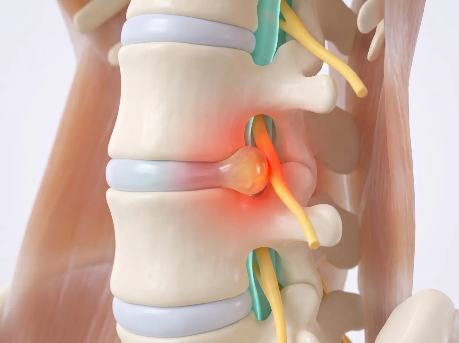
Often driven by a disc or bone spur pressing on a nerve root
Calmed by taking pressure OFF the nerve: positional exercises, gentle nerve glides, and epidural injections
Joint pain — it stays put
Aches right at the joint; stiff in the morning, sore after loading
Feels deep and local — it does not race down the limb
Driven by cartilage wear and joint inflammation (arthritis)
Helped by strengthening AROUND the joint, steady movement, and targeted joint injections or nerve ablation
A surprising factUp to 4 in 10 cases of isolated arm or leg pain actually start at the spine — which is why treating only the shoulder, elbow, or knee sometimes fails until the neck or back is addressed.
The direction ruleWith nerve pain, the right exercise makes pain retreat from the limb back toward the spine (“centralizing”) — a good sign. Pain spreading further down the limb means stop that movement.
Slides, not pullsIrritated nerves hate hard stretching. Nerve exercises are gentle rhythmic slides that help the nerve move and drain swelling — never forceful pulls.
Joints like loadWorn joints do best with the opposite: progressively stronger muscles around them to absorb force — motion is lotion, and strength is the shock absorber.
Not sure which type you have — or have some of both? That is exactly what your evaluation sorts out, using your story, your exam, and how your pain responds to movement (imaging alone often cannot tell: many pain-free people have “abnormal” scans).
🦴 Neck procedures
Procedures for pain that starts in the cervical spine — neck pain, stiffness, headaches from the neck, and nerve pain that radiates into the shoulder or arm.
Cervical Epidural Steroid Injection
Relief for nerve pain that runs into the shoulder or arm
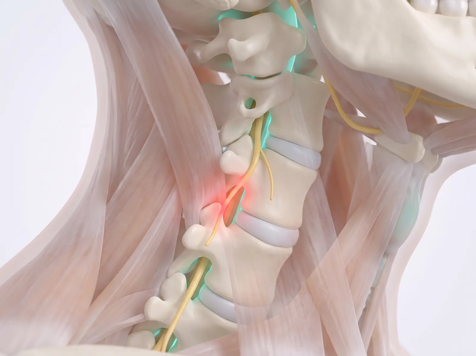
An image-guided injection that places anti-inflammatory medicine into the epidural space of the neck — the protective sleeve just outside the sac that surrounds the spinal cord and nerve roots.
Often used for: Pinched nerves in the neck from a herniated disc or arthritis narrowing — pain, tingling, or numbness radiating to the shoulder blade, arm, or hand.
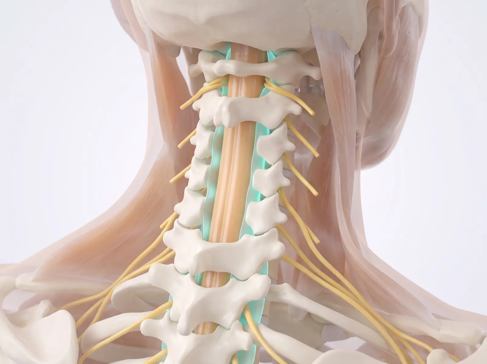
Seven small vertebrae protect the spinal cord as it carries signals between brain and body. Nerve roots exit at each level toward the shoulders and arms; the epidural space (teal) surrounds and cushions them.
2Where the pain starts
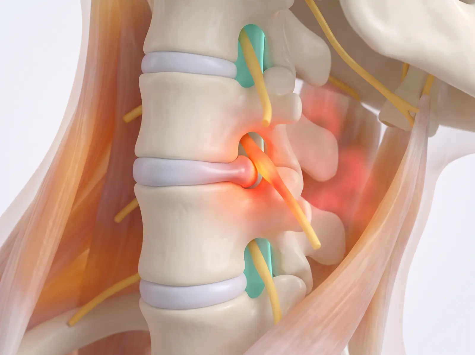
A worn or herniated disc can press on an exiting nerve root and inflame it — causing pain, tingling, or numbness that travels along that nerve into the arm.
3The procedure
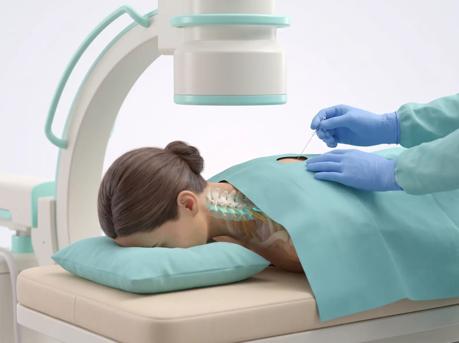
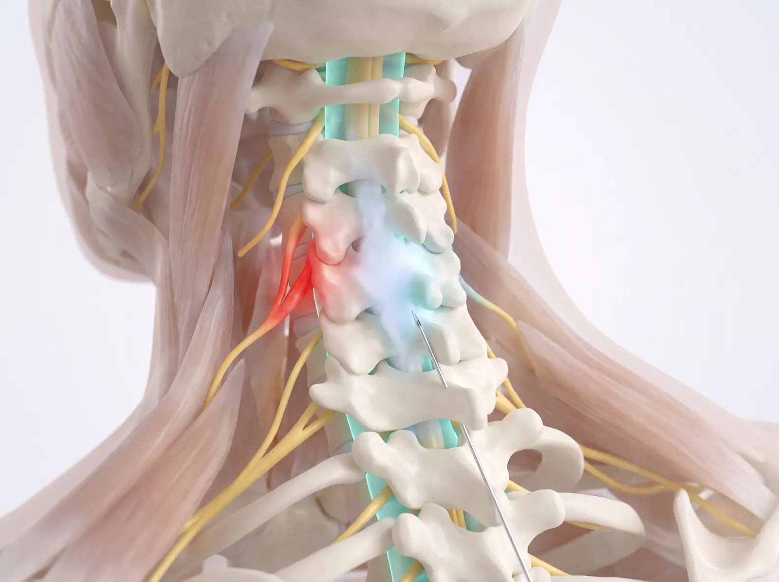
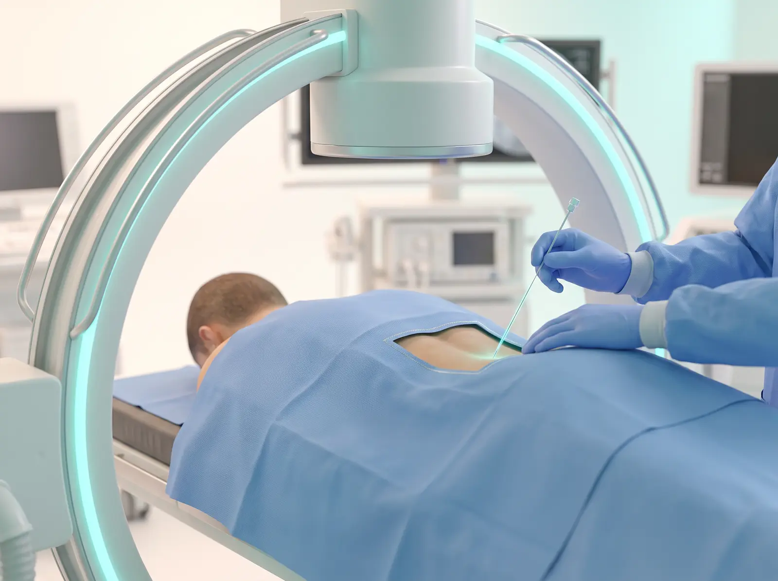
You lie face down with your forehead supported. Skin is numbed first. Using continuous X-ray guidance, a very thin needle is advanced at the base of the neck — the widest, safest entry level.
4Relief delivered
The needle tip stops in the epidural space, outside the nerve sac — it never touches the spinal cord. Anti-inflammatory medicine spreads gently upward and bathes the irritated root, cooling the inflammation so it can settle.
What to expect: Mild soreness for a day or two is normal. Relief usually builds over 3–7 days as the steroid takes effect.
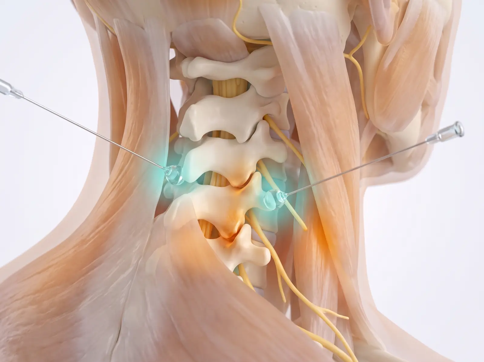
Finding — then switching off — arthritic joint pain in the neck
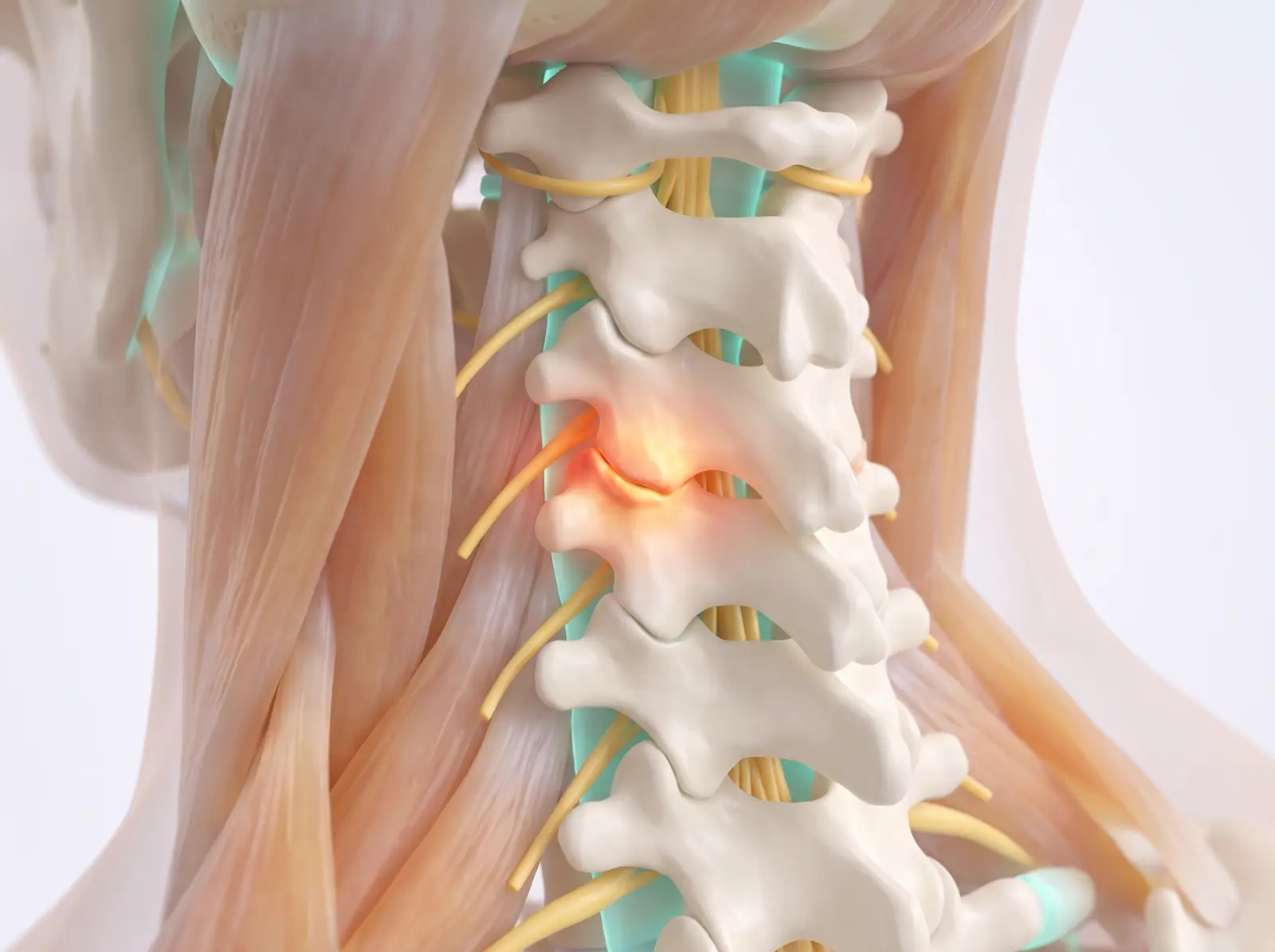
Each facet joint of the neck reports its pain through two tiny nerves called medial branches. This two-step approach first tests whether those joints are truly the source, then quiets the confirmed nerves with precise heat.
Often used for: Neck pain and stiffness from facet-joint arthritis — often worse looking up or turning, sometimes with headaches that start at the base of the skull.
Facet joints are the small joints at the back of the spine where each vertebra glides on the next. With arthritis they become inflamed (shown glowing) and send steady aching signals through their tiny medial branch nerves.
2Step one — the test block
Through very thin needles, a drop of numbing medicine is placed precisely where each medial branch crosses the bone. If your usual pain drops substantially while the numbing lasts, the joints are confirmed as the source.
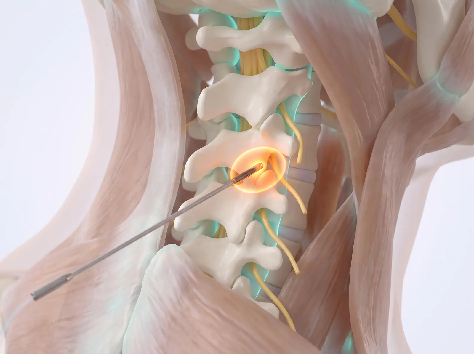
3Step two — radiofrequency ablation
A slender cannula with a heated tip is placed alongside each confirmed nerve. It creates one small, controlled heat lesion — just millimeters wide — interrupting the pain signal without disturbing surrounding structures.
4Lasting relief
The treated nerves can no longer deliver the joint’s pain message. Relief typically lasts 9–18 months. The nerves slowly regrow, and the procedure can be safely repeated.
What to expect: A test block wears off the same day — that is expected; it is a diagnostic. After ablation, soreness for up to a week, then relief settles in over 2–4 weeks.
🧍 Low back & pelvis procedures
Procedures for low-back pain, buttock pain, and sciatica — from the lumbar spine, its facet joints, and the sacroiliac joints where the spine meets the pelvis.
Lumbar Epidural Steroid Injection
Calming an inflamed nerve in the low back
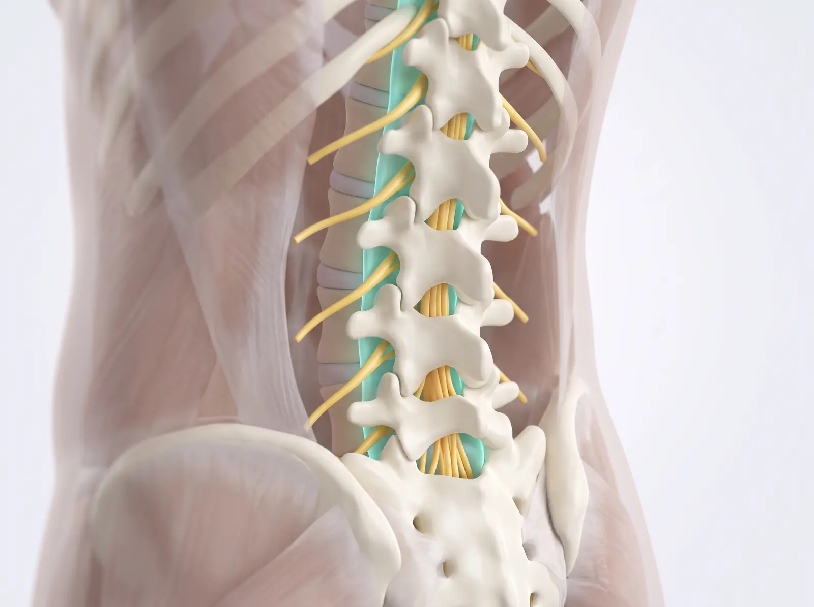
An image-guided injection that places anti-inflammatory medicine into the epidural space — the cushioned sleeve just outside the sac that protects your spinal nerves.
Often used for: Sciatica — leg pain, numbness, or tingling from a herniated disc or spinal stenosis — often alongside low-back pain.
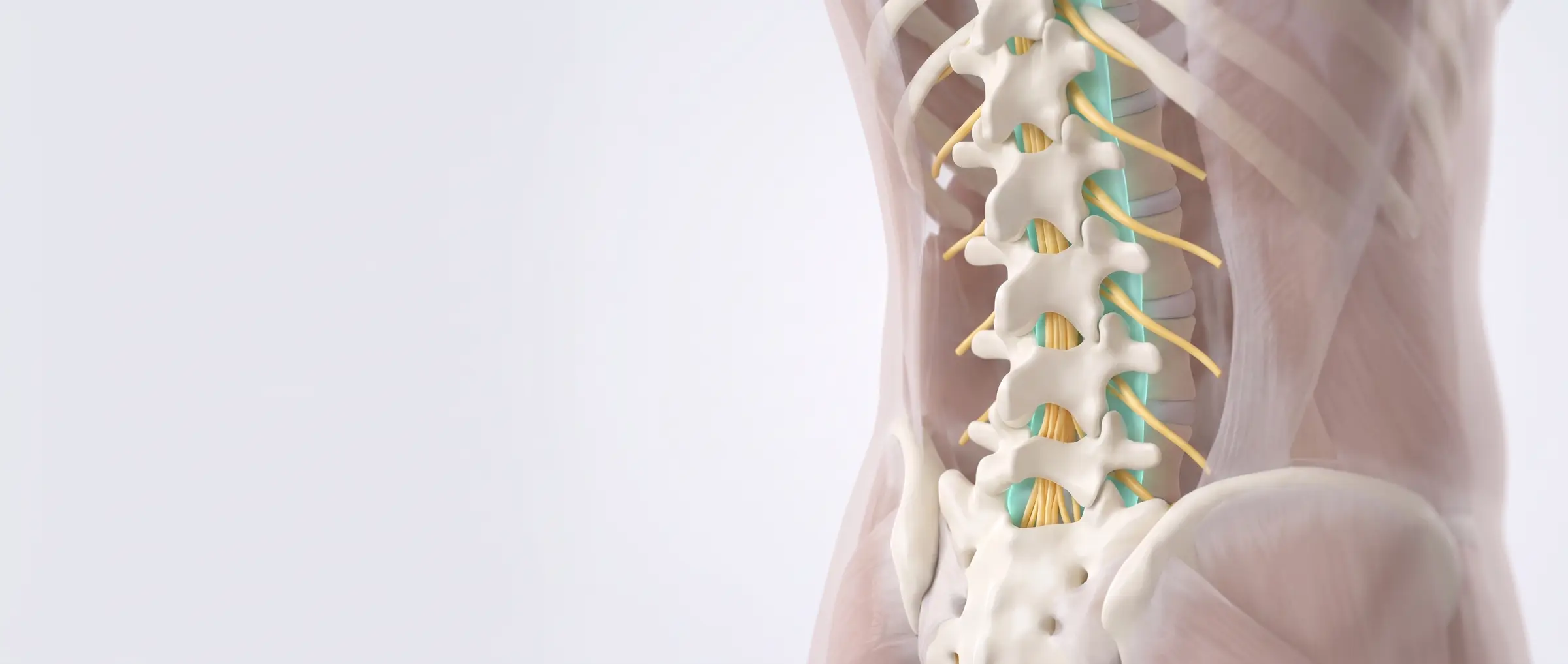
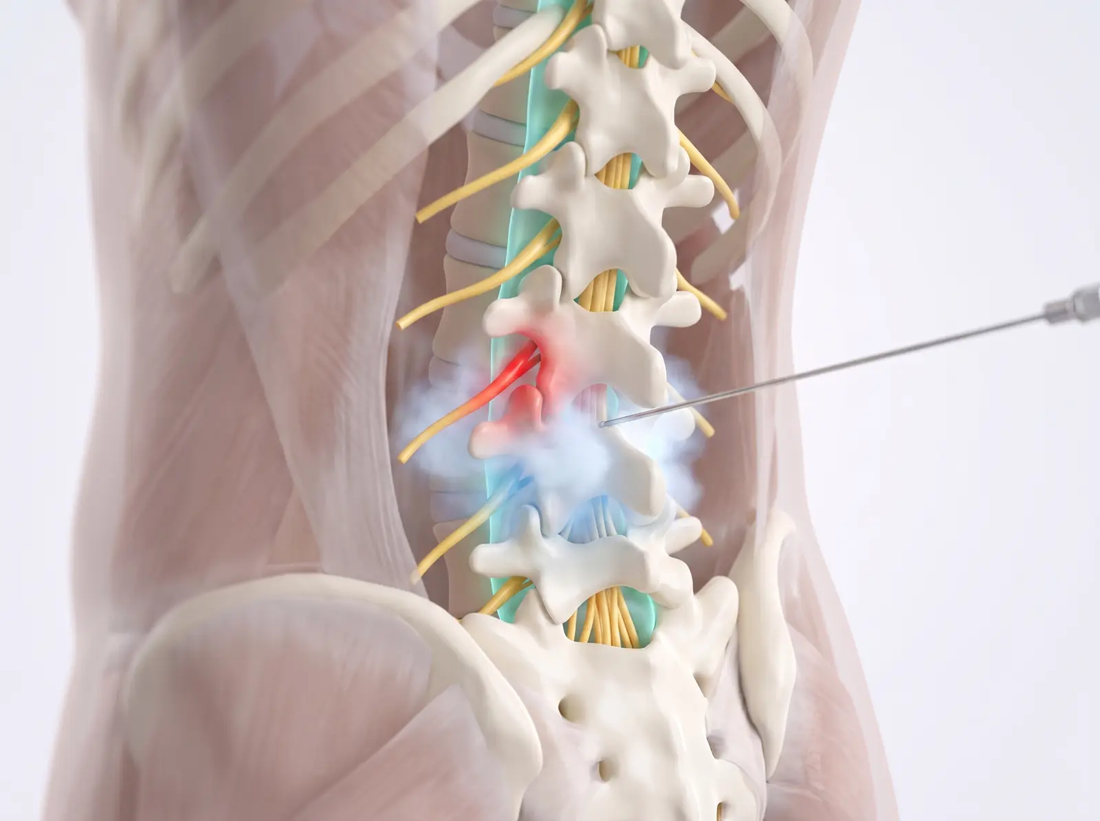
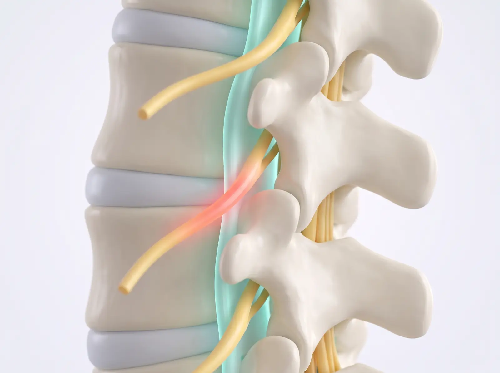
The nerves of the low back travel inside the spinal canal and exit between the vertebrae on their way to the legs. The epidural space (teal) surrounds and cushions them.
2Where the pain starts
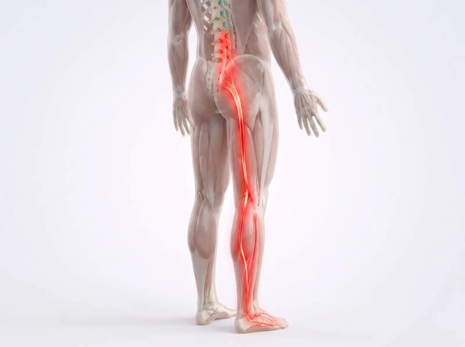
A bulging or herniated disc can press on a nerve root and inflame it. That irritation is what sends pain shooting down the leg — what most people call sciatica.
3The procedure
You lie face down while a low-dose X-ray camera shows the exact path. After numbing the skin, a very thin needle is guided between the vertebrae. Most patients feel pressure more than pain; the injection itself takes only a few minutes.
4Relief delivered
The needle tip stops in the epidural space — outside the nerve sac. A mix of steroid and anesthetic bathes the inflamed root, cooling the irritation so the nerve can settle and heal.
What to expect: You go home the same day and can usually return to normal activity tomorrow. Relief often builds over 3–7 days.
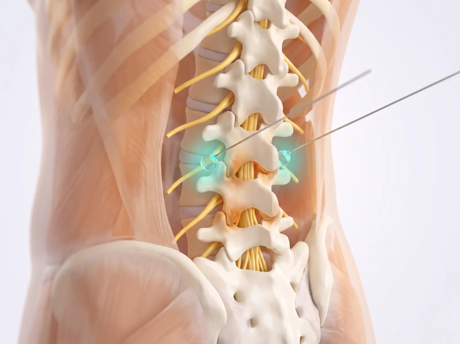
Finding — then switching off — arthritic joint pain in the back
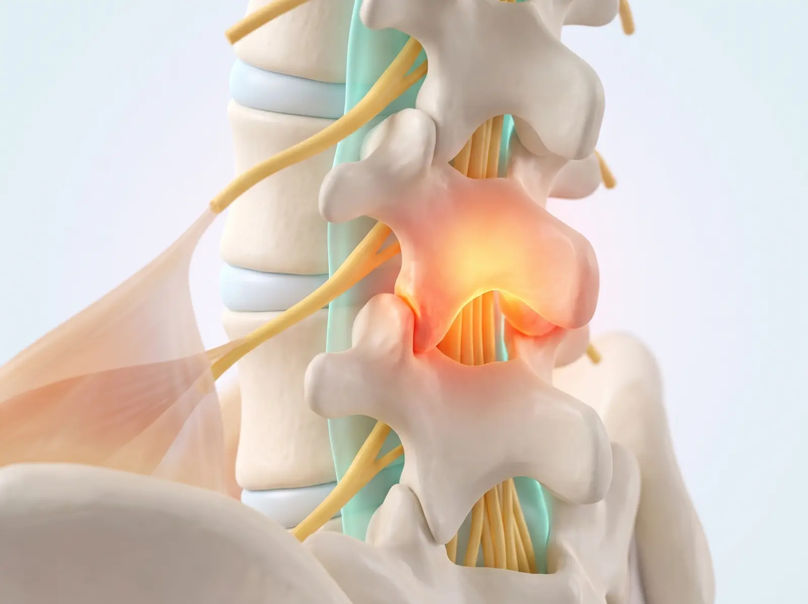
Each facet joint of the low back reports its pain through two tiny medial branch nerves. First a numbing test confirms the joints are the real source; then radiofrequency heat quiets those nerves for long-lasting relief.
Often used for: Back pain from facet-joint arthritis — typically worse with standing, arching backward, or twisting, and better sitting.
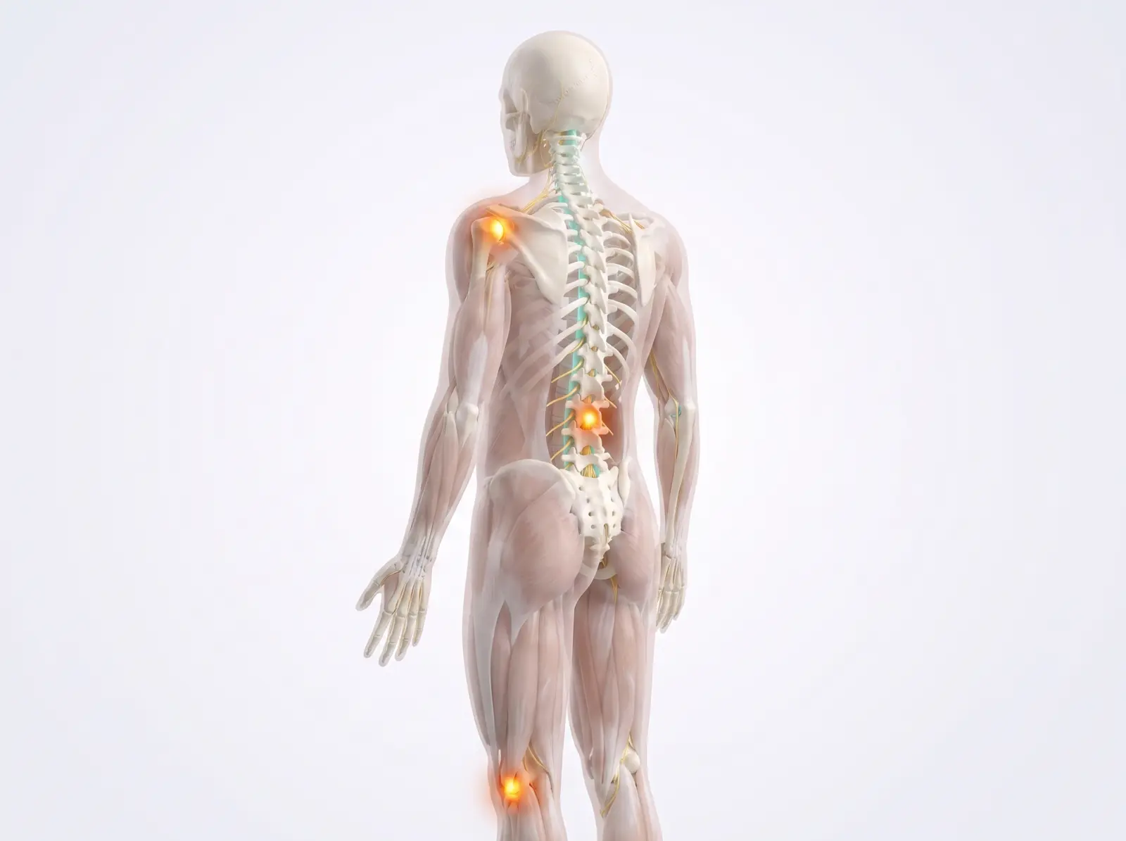
Facet joints are the paired joints where each vertebra interlocks with the next. Arthritic ones become inflamed (shown glowing) and ache — the signal carried by two tiny medial branch nerves per joint.
2Step one — the test block
Very thin needles place a drop of numbing medicine exactly where each medial branch crosses the bone. If your usual pain drops substantially for the next hours, the joints are confirmed as the pain source.
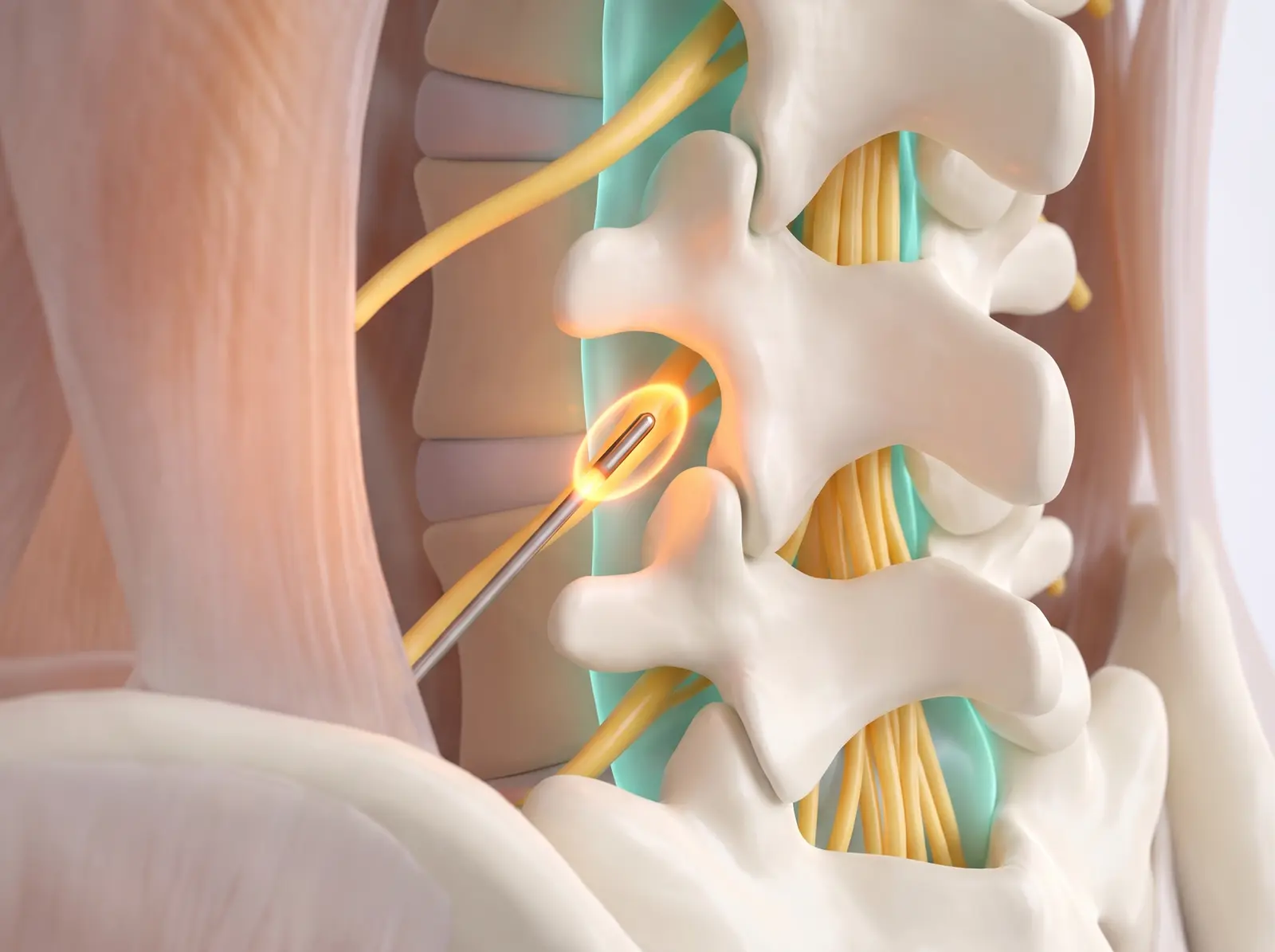
3Step two — radiofrequency ablation
A slender heated tip lies alongside each confirmed nerve and creates a precise, millimeters-wide heat lesion — interrupting the pain signal while everything around it is protected.
4Lasting relief
With its messenger nerves quieted, the joint’s pain message no longer reaches the spine. Relief typically lasts 9–18 months, and the procedure can be repeated when the nerves regrow.
What to expect: After ablation expect up to a week of soreness (like a deep bruise), then steadily improving relief over 2–4 weeks.
Sacroiliac (SI) Joint Injection
Treating the joint where your spine meets your pelvis
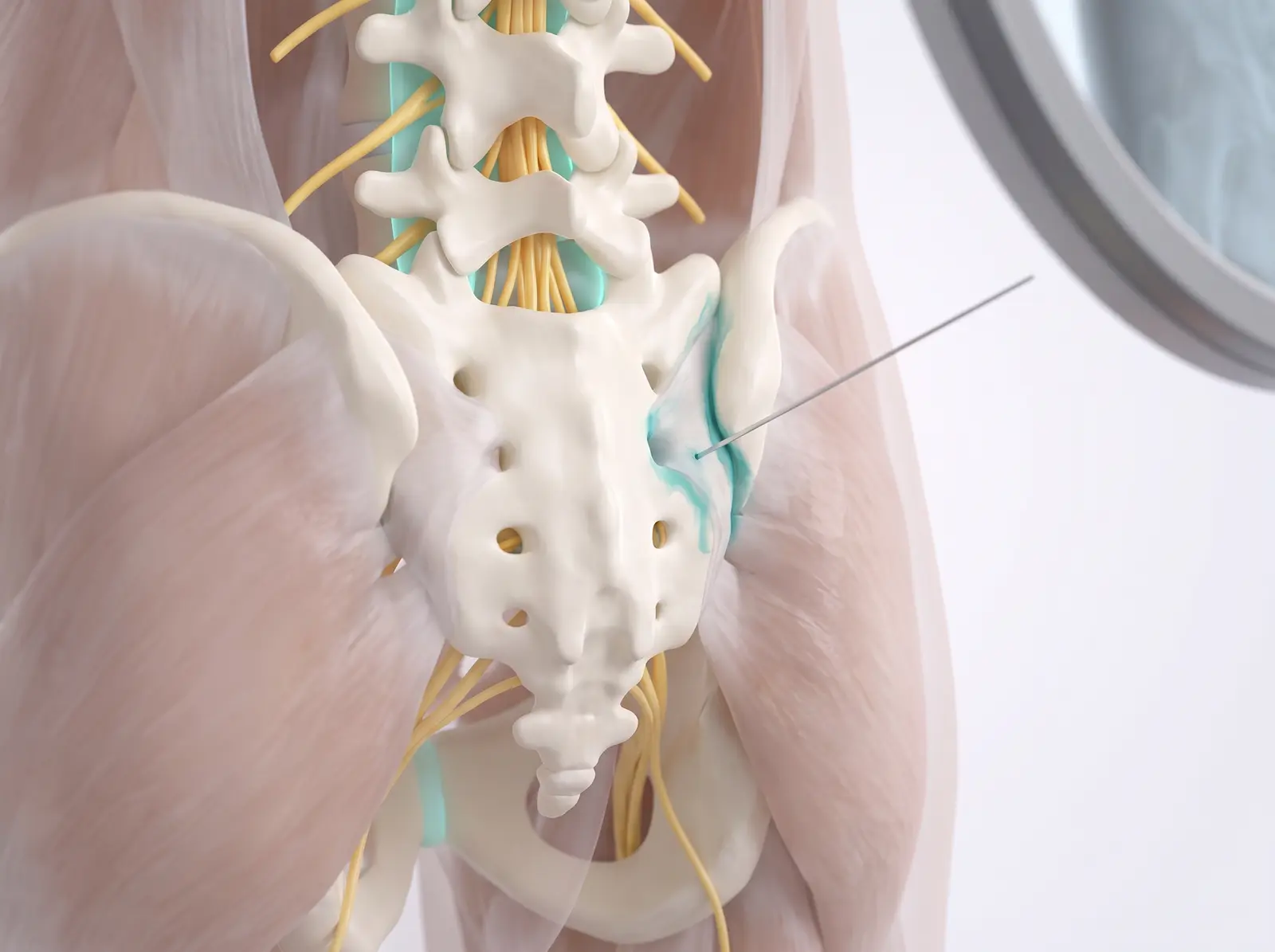
An image-guided injection of anti-inflammatory medicine directly into the sacroiliac joint — the strong, ligament-wrapped joint connecting the base of the spine to the pelvis.
Often used for: One-sided low-back and buttock pain — often felt standing up from a chair, climbing stairs, or rolling over in bed.
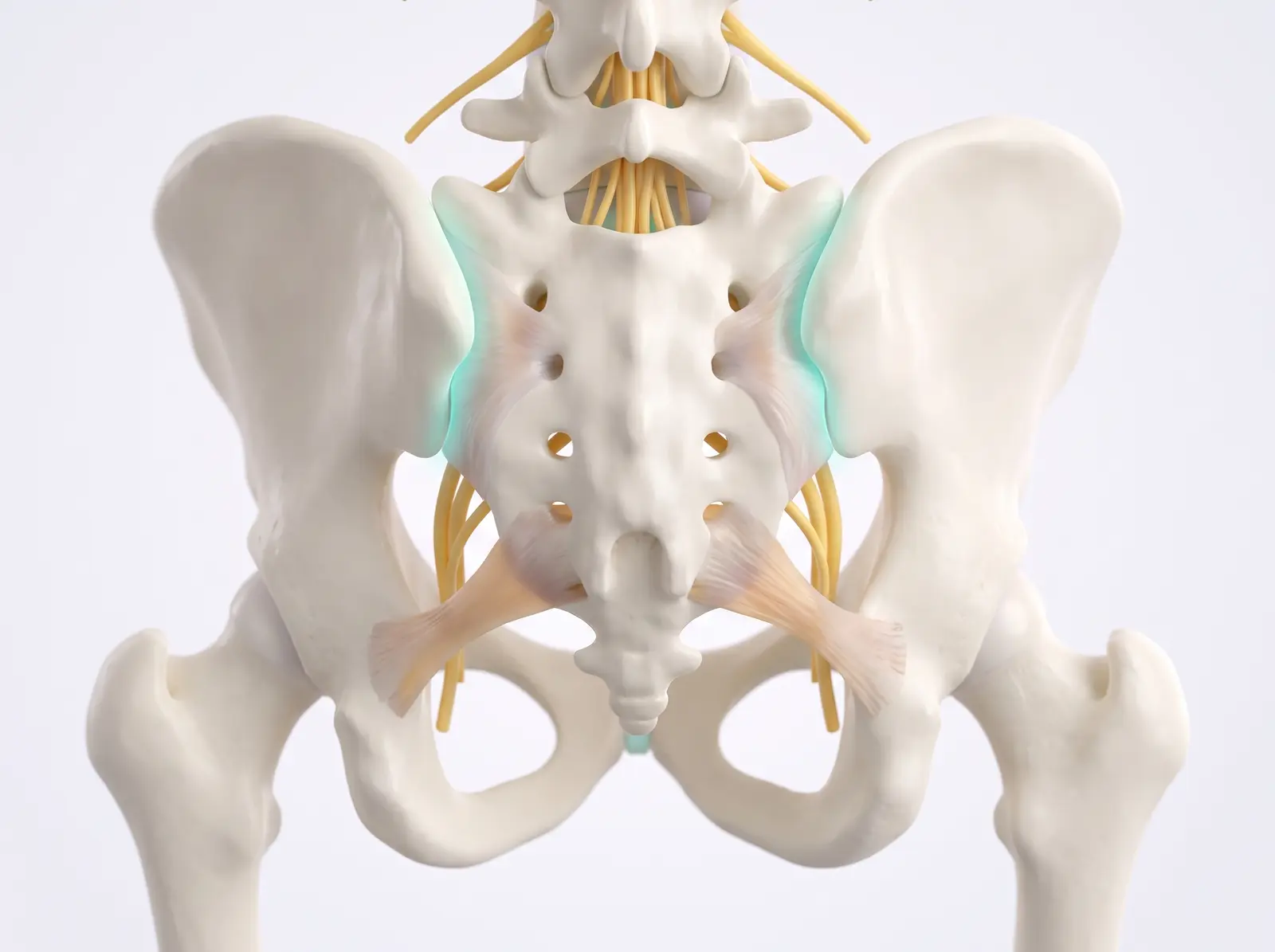
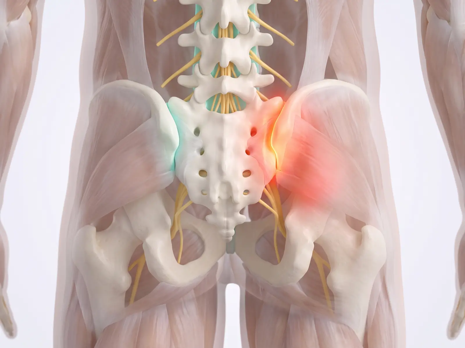
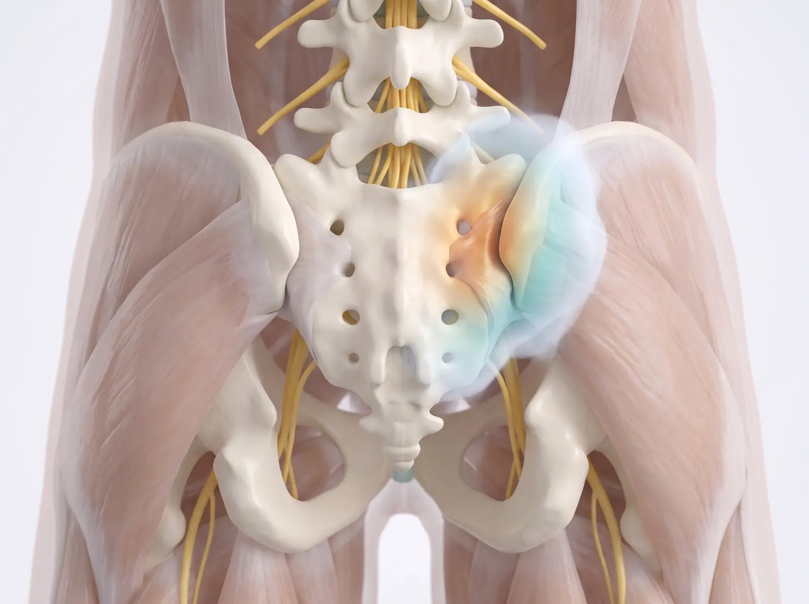
The two sacroiliac joints connect the sacrum — the base of your spine — to the pelvis. Crossed by some of the strongest ligaments in the body, they transfer your upper-body weight into the legs with every step.
2Where the pain starts
When an SI joint becomes inflamed or moves poorly, it produces one-sided pain over the buttock (shown glowing), sometimes spreading toward the back of the thigh.
3The procedure
Under X-ray guidance, a thin needle enters the lower part of the joint. A whisper of contrast dye outlines the joint line, confirming the medicine will reach exactly the right spot.
4Relief delivered
Anti-inflammatory medication fills the joint and calms the inflammation. The injection is diagnostic too — if the pain eases, the SI joint is confirmed as the true source.
What to expect: Home the same day. Numbing medicine may give hours of early relief; the steroid effect builds over the following week.
⚡ Nerve stimulation
For chronic nerve pain that hasn’t responded to medications, therapy, or injections — technology that intercepts pain signals on their way to the brain.
Spinal Cord Stimulation — Trial, then Implant
A pacemaker for pain — always test-driven first
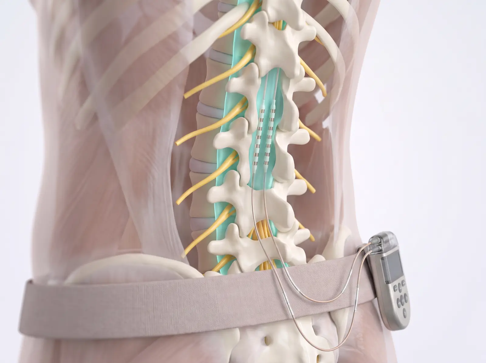
A spinal cord stimulator delivers gentle electrical pulses to the spinal cord that mask pain signals before they reach the brain. It is always tried first with temporary leads — no incision — so you can test the relief in your own daily life.
Often used for: Persistent nerve pain — after back surgery, from neuropathy, or complex regional pain — when conservative care hasn’t been enough.
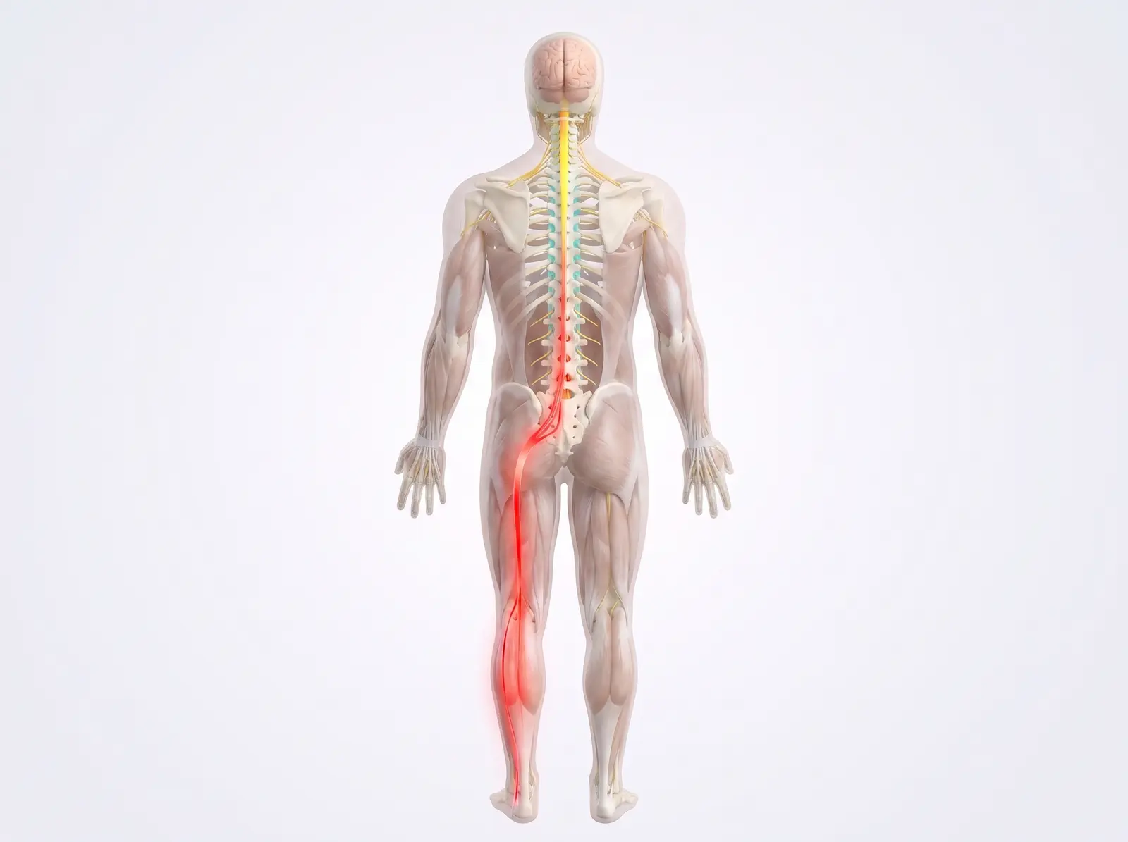
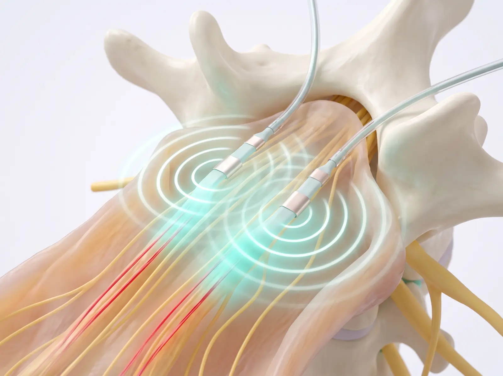
Chronic nerve pain travels from the body up the spinal cord to the brain (shown in red). If the message can be softened on the way up, the brain simply receives less pain.
2The trial — no incision
Through a needle, thin temporary leads are threaded into the epidural space over the spinal cord and connected to a small external stimulator worn on a belt. You go home the same day and live your normal week.
3How it works
The leads deliver gentle electrical pulses that scramble or mask the ascending pain signal — many patients feel a soft tingling, or simply notice the pain fading into the background.
4The implant
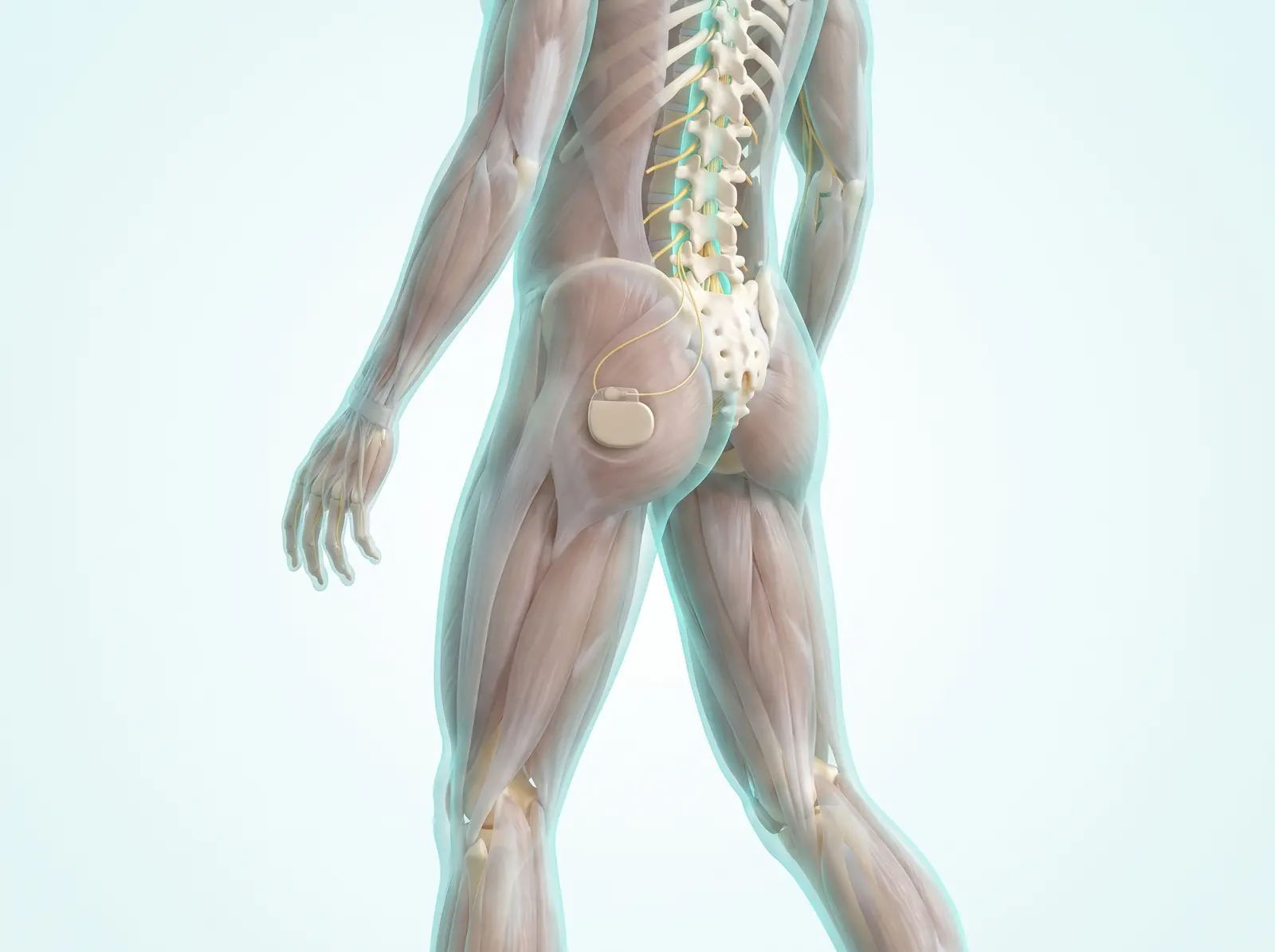
If the trial week gives meaningful relief — typically at least half your pain — a permanent system with a small battery under the skin, much like a pacemaker, can be implanted. You adjust it with a handheld remote, and it can be turned off or removed.
What to expect: The trial is the whole point: you judge the benefit yourself, at home, before any implant decision is made.
🏃 Home exercise library
One position per photo — pairs show the start and end of a movement, and the ▶ videos demonstrate the full motion. Each exercise is tagged for its target: Nerve-calming gentle glides and decompression positions, or Joint & muscle strengthening and mobility. Move slowly, breathe, and stay within comfortable limits. 🖨 Print the full exercise library.
🧘 Low back
Leg pain that shoots below the knee is usually NERVE-type — start with the McKenzie press-up progression and sciatic slider, and watch the direction rule. Local back ache with stiffness is usually JOINT-type — build up the stabilizers.
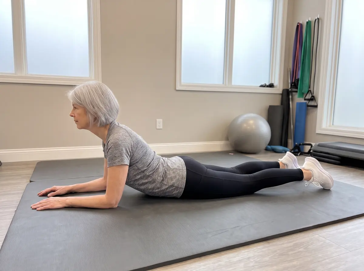
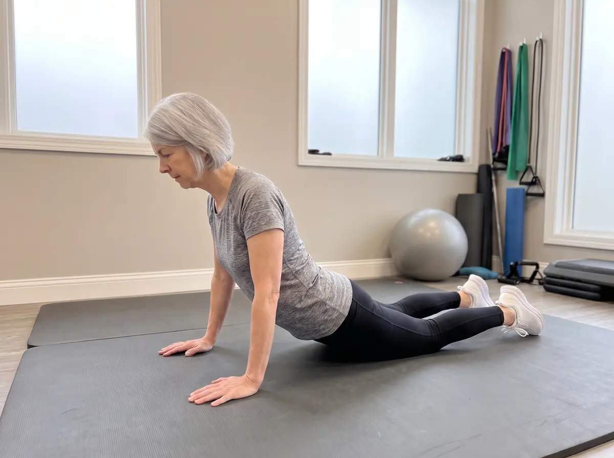
1 · On elbows2 · Press-up
▶ Watch the movement
McKenzie Extension ProgressionNerve-calming
Lie face down 2–3 minutes, prop onto elbows 2–3 minutes, then press the chest up with hips staying on the mat — 10 gentle reps. If your leg pain retreats toward the spine, this is your exercise; if it spreads further down the leg, stop.
PointFlex
▶ Watch the movement
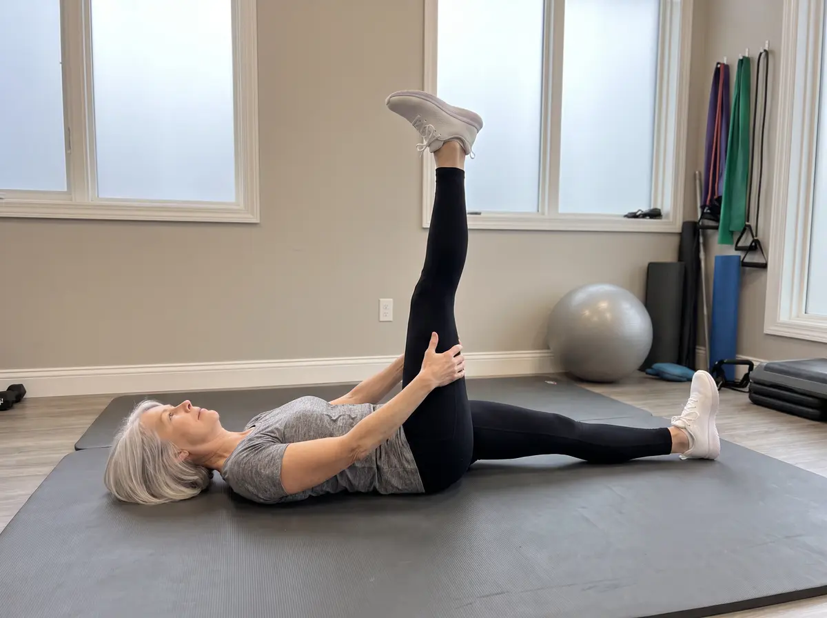
Sciatic Nerve SliderNerve-calming
Holding behind the thigh, alternate smoothly: straighten the knee with the foot pointed — then bend the knee and pull the foot up. A gentle rhythmic slide, never a hard stretch. 10–15 slow cycles per side.
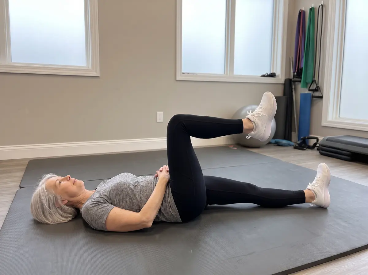
Single Knee to ChestNerve-calming
For stenosis-type pain (worse standing, better sitting): pull one knee toward the chest until an easy stretch. Hold 20–30 seconds; 3 times each side.
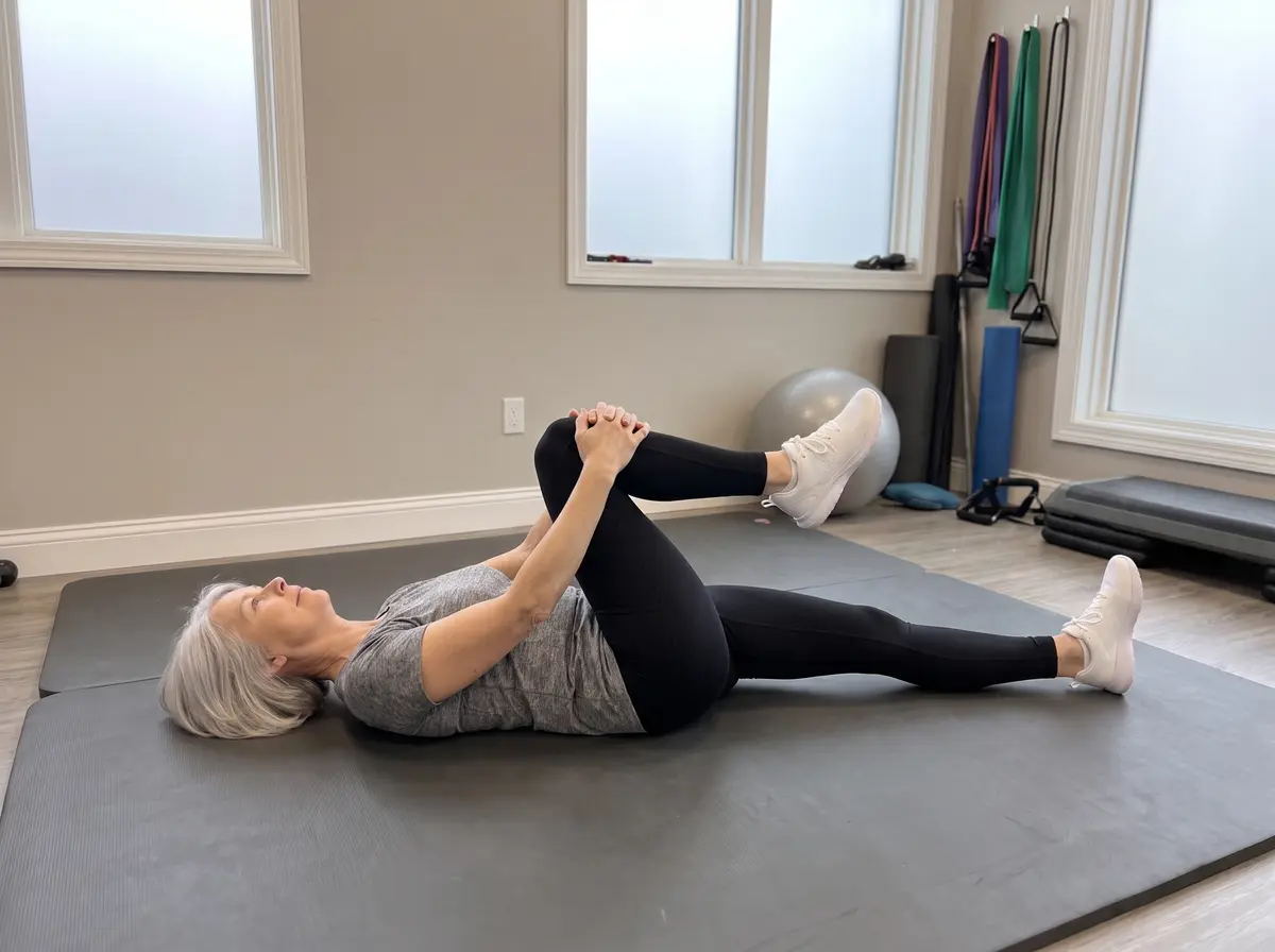
Double Knee to ChestNerve-calming
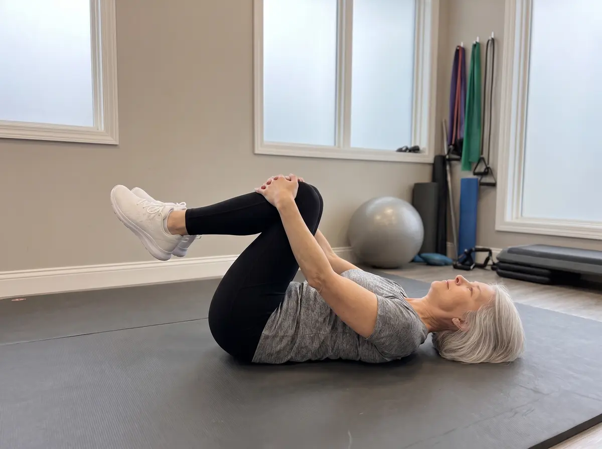
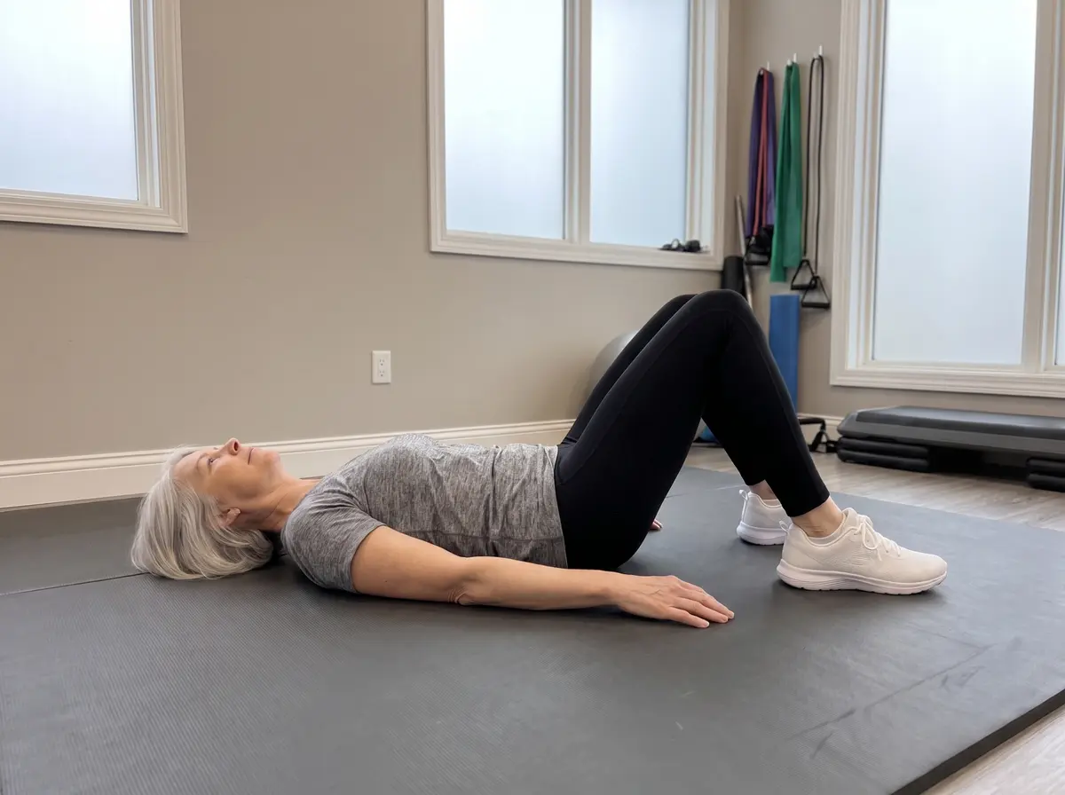
Hug both knees toward the chest, letting the low back round gently — this opens the nerve passageways. Hold 20–30 seconds; repeat 3 times.
CatCow
▶ Watch the movement
Cat – CowJoint & muscle
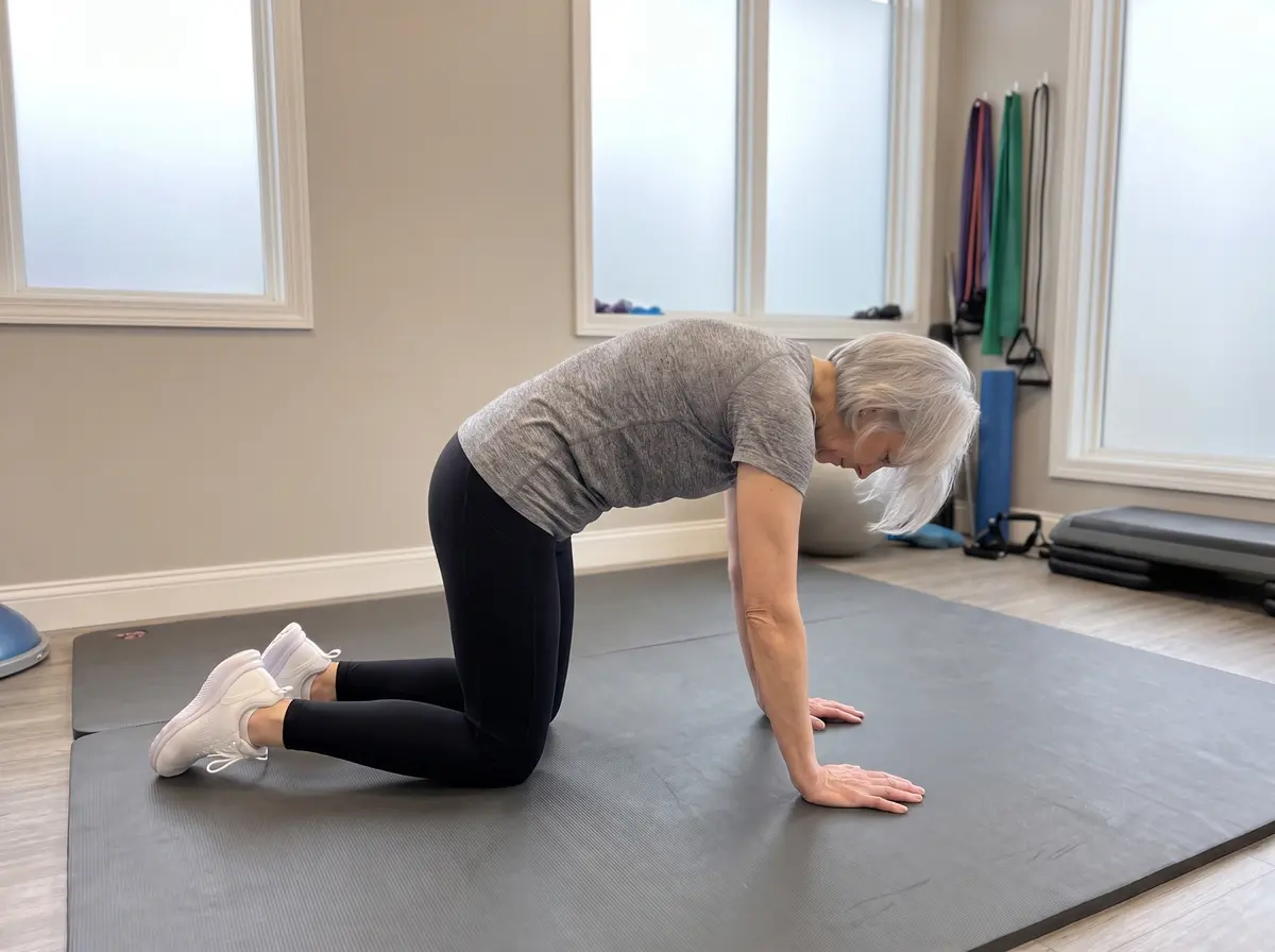
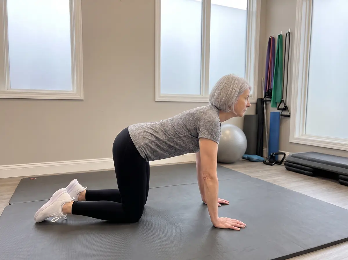
Round your back up like a cat, then let the belly sink while the chest lifts. Move slowly with your breath. 10 cycles.
▶ Watch the movement
Bird-DogJoint & muscle
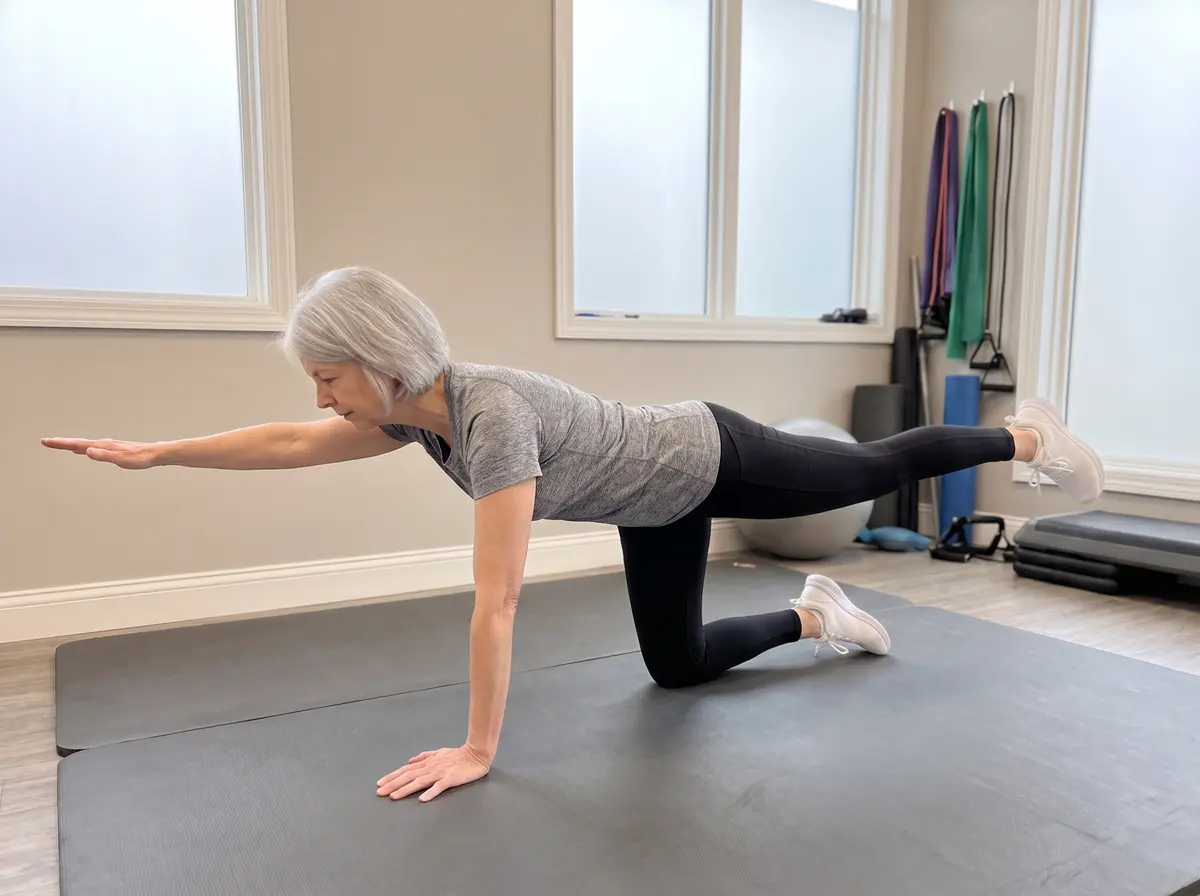
Reach one arm forward and the opposite leg back until level. Hips square, back flat — this trains the deep stabilizers that protect arthritic segments. Hold 5 seconds; 10 per side.
StartLift
▶ Watch the movement
BridgeJoint & muscle
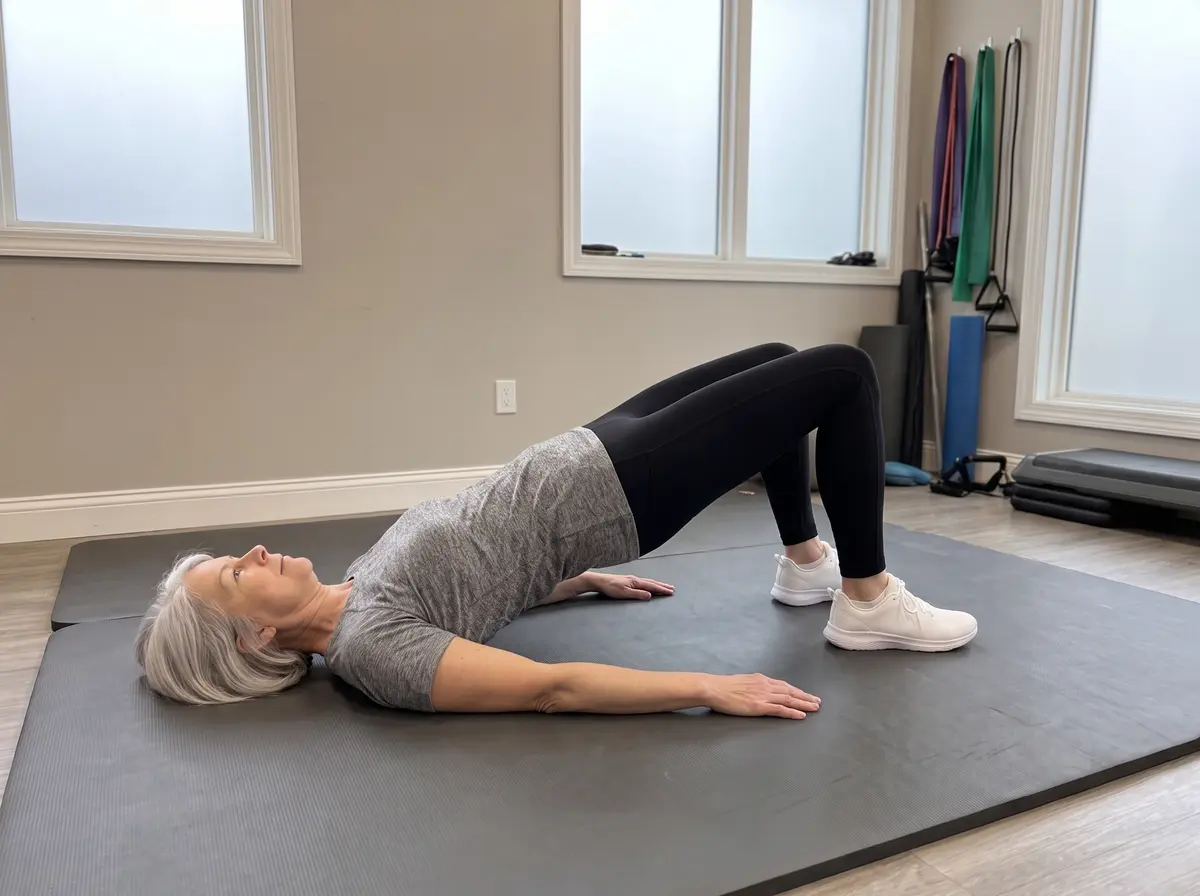
Squeeze the buttocks and lift the hips until shoulders, hips and knees form a line. Hold 5 seconds, lower with control. 10 repetitions.
Child’s PoseJoint & muscle
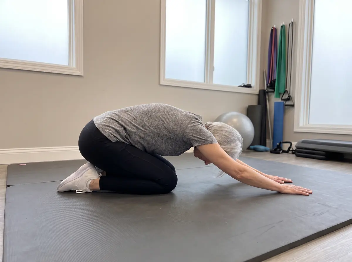
Sit back onto your heels and reach forward, letting the back lengthen. A resting position between sets. Hold 30 seconds; 3 times.
🙆 Neck
Arm pain, tingling, or numbness points to a NERVE target — chin tucks and nerve sliders, guided by the direction rule. Local neck ache and stiffness points to the JOINT target — deep-flexor control and gentle stretches.
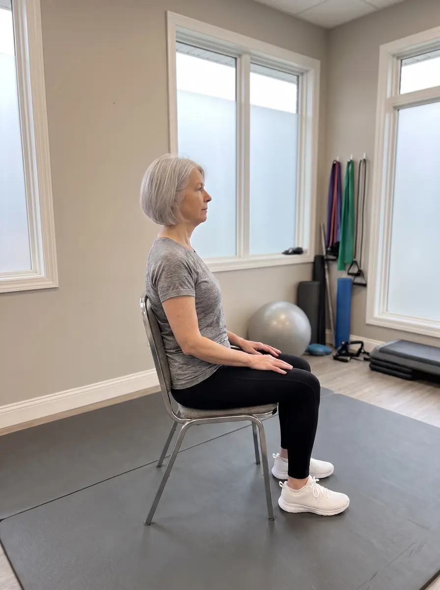
StartTuck
▶ Watch the movement
Chin Tuck (Retraction)Nerve-calming
Glide the chin straight back — a gentle double chin — eyes level. This decompresses the lower neck where pinched nerves live. Hold 5 seconds; 10 repetitions. Arm pain easing toward the neck = the right direction.
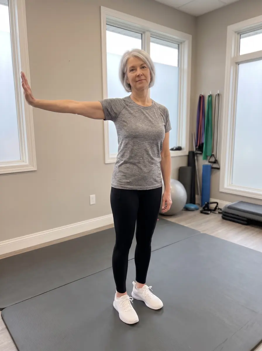
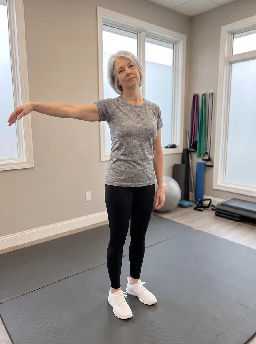
Wrist back · head towardWrist easy · head away
Median Nerve SliderNerve-calming
Arm out at shoulder height. As the wrist bends back, tilt your head TOWARD that arm; as the wrist relaxes, tilt AWAY. The nerve glides without being yanked. 10–15 smooth cycles. Mild tingling that fades is fine; electric pain is not.
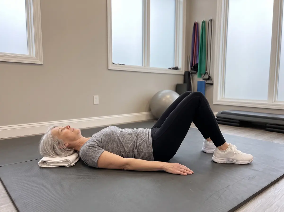
Deep Neck Flexor NodJoint & muscle
Lying with knees bent, nod the chin gently toward the throat — a tiny “yes” — without lifting the head. This wakes the deep stabilizers that support arthritic segments. Hold 10 seconds; 10 reps.
Levator StretchJoint & muscle
Turn the head 45° and look down toward your pocket; the hand adds gentle weight. Hold 20–30 seconds; 3 times each side.
Upper Trapezius StretchJoint & muscle
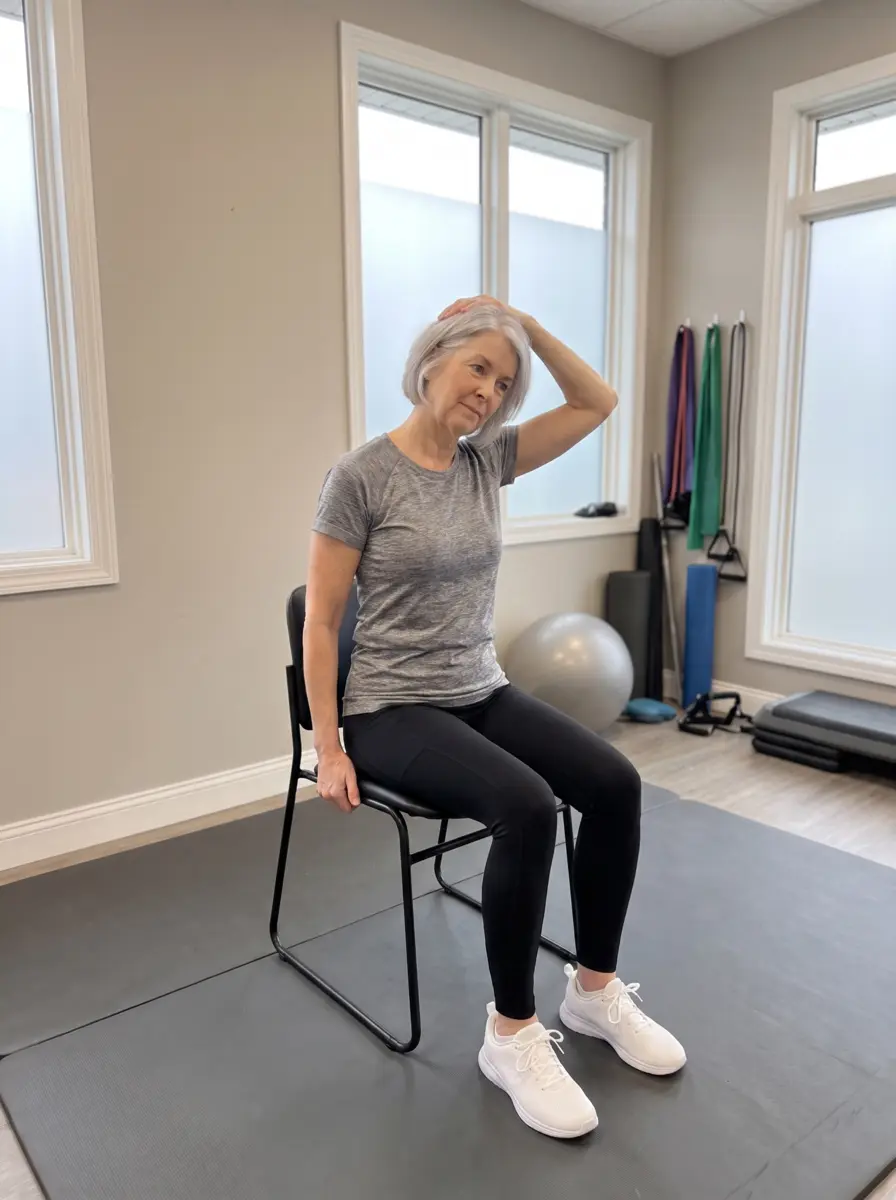
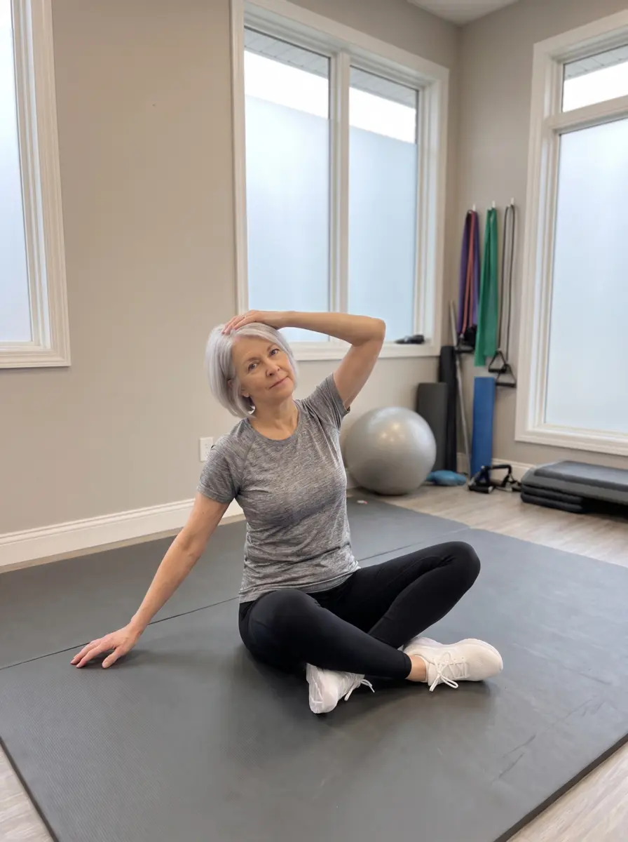
Ear toward one shoulder, gentle overpressure, other shoulder relaxed and low. Hold 20–30 seconds; 3 times each side.
Neck RotationJoint & muscle
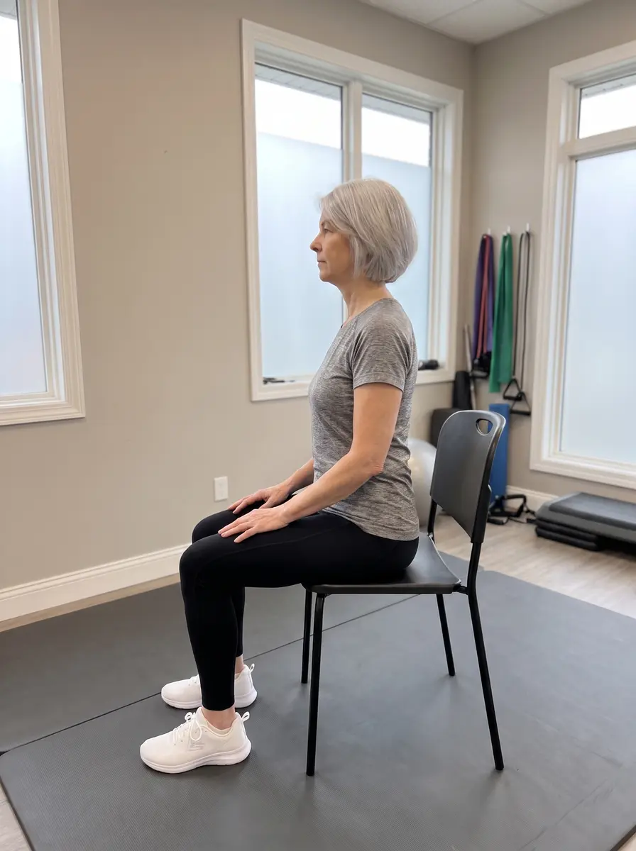
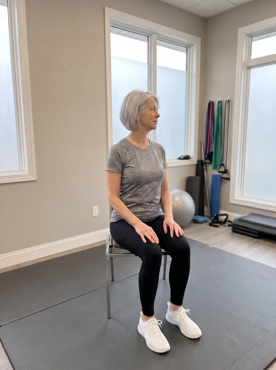
Sitting tall, slowly look over one shoulder as far as comfortable. Hold 10–15 seconds; 5 times each direction.
🔗 Mid-back — the connector
The mid-back is the stiffest part of the spine. When it stops extending, the neck and low back are forced to over-move — so freeing it protects BOTH neighbors. These help nearly everyone.
Seated Thoracic ExtensionFor everyone
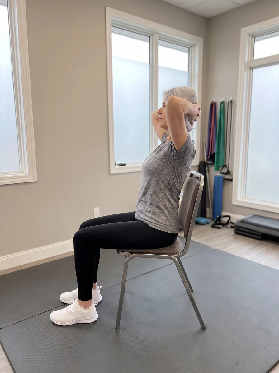
Hands behind the head, arch the upper back over the top of the chair’s backrest, chest to the ceiling. 10 slow arches — feel it between the shoulder blades, not in the low back.
Open BookFor everyone
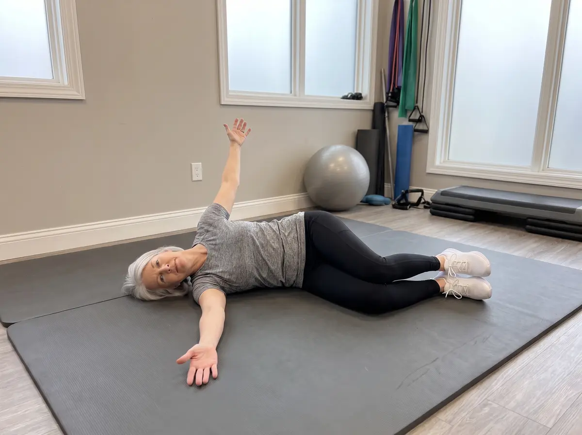
Side-lying with knees stacked, sweep the top arm open like a book, chest rotating to the ceiling, eyes following the hand. 8–10 slow openings per side.
💪 Shoulder & shoulder blade
Most shoulder pain improves when the shoulder BLADE muscles fire correctly again — that is what the Y, T, W and push-up plus retrain. If your “shoulder pain” comes with neck symptoms or tingling below the elbow, work on the neck first.
Prone “Y”Joint & muscle
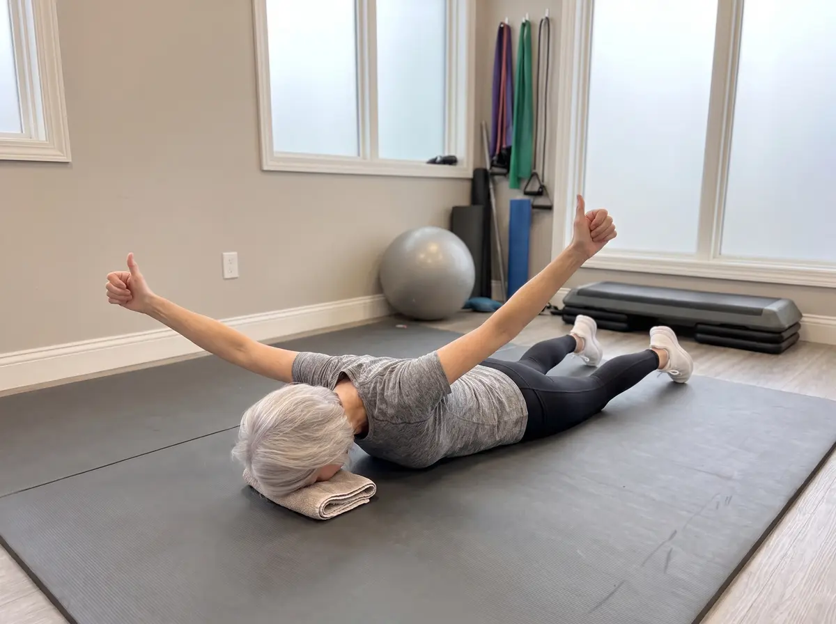
Face down, arms overhead in a Y, thumbs up. Lift the arms a few inches by drawing the shoulder blades down and in — the single best lower-trapezius builder. Hold 3 seconds; 10 reps.
Prone “T”Joint & muscle
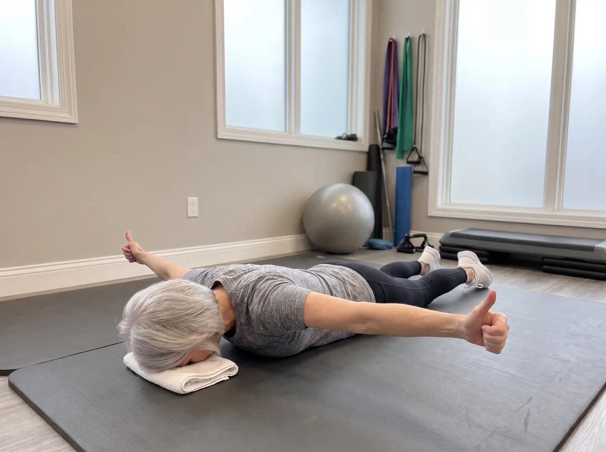
Arms straight out in a T, thumbs up. Squeeze the shoulder blades together as the arms lift. Hold 3 seconds; 10 reps.
Prone “W”Joint & muscle
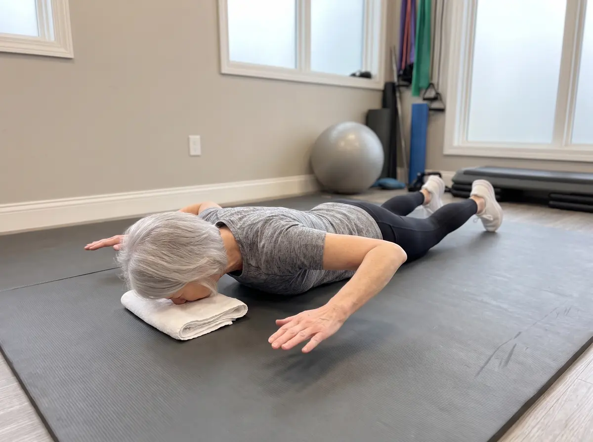
Elbows bent in a W, draw the elbows down and back toward the hips, blades gliding down and together. Hold 3 seconds; 10 reps.
Push-Up PlusJoint & muscle
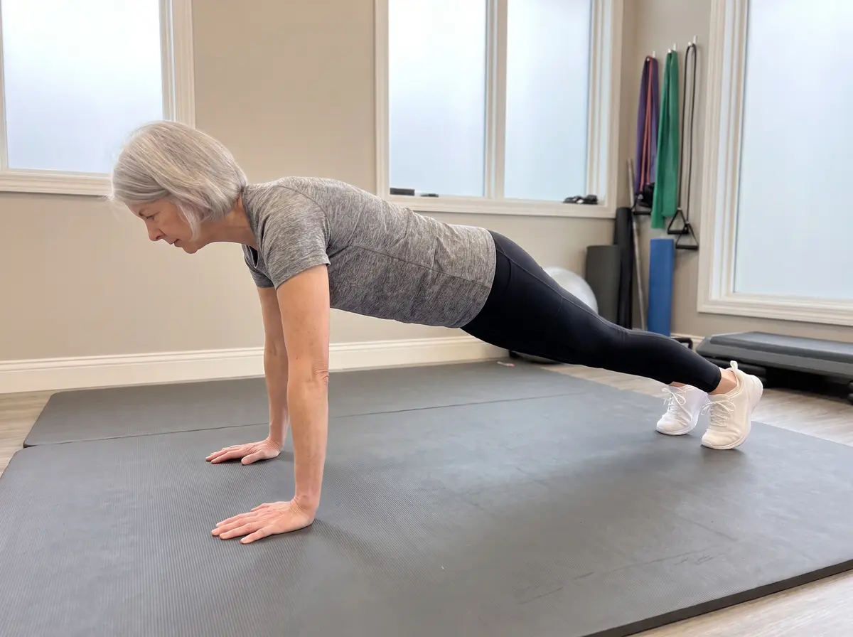
At the top of a push-up (knees down is fine), keep the arms straight and push the floor away an extra inch so the upper back rounds slightly — this fires the serratus anterior that steers the shoulder blade. 10 slow reps.
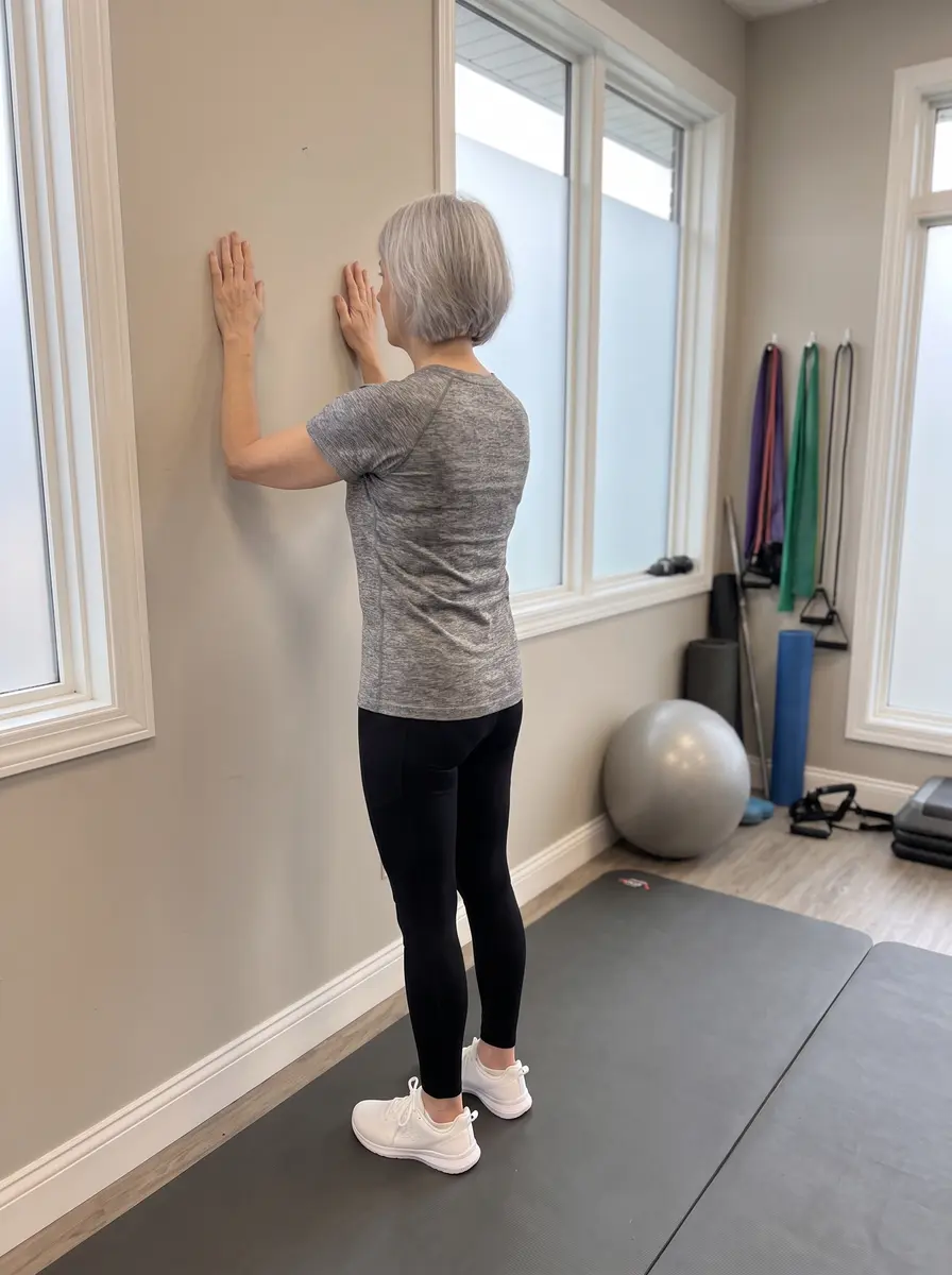
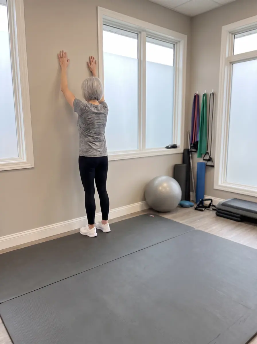
StartSlide up
▶ Watch the movement
Wall SlideJoint & muscle
Forearms on the wall at shoulder height, slide up as high as comfortable, shoulders relaxed, then lower. 10 slow repetitions.
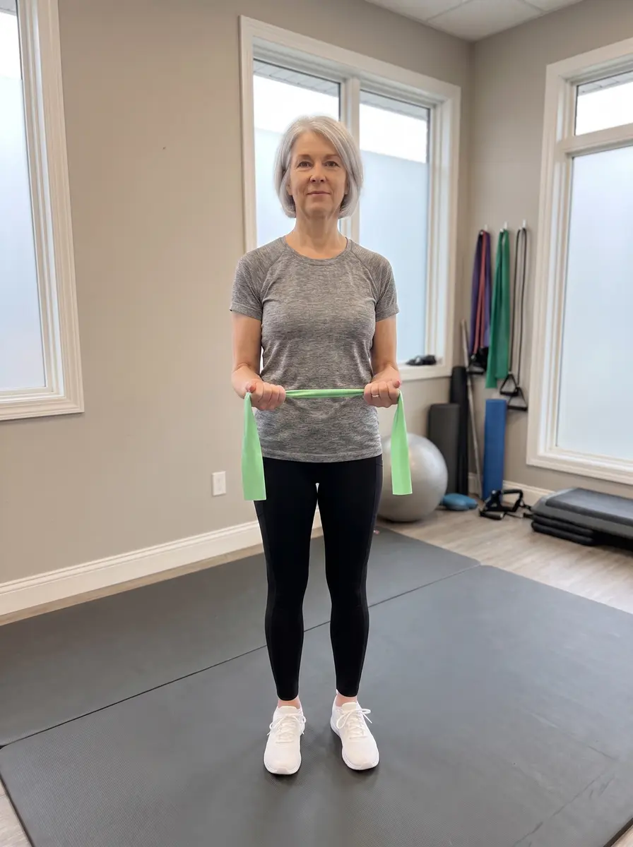
StartRotate out
▶ Watch the movement
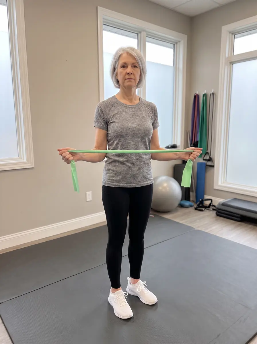
Band External RotationJoint & muscle
Elbows bent 90° and pinned to your sides, rotate the forearms outward against the band. Slow out, slow back. 10–15 repetitions.
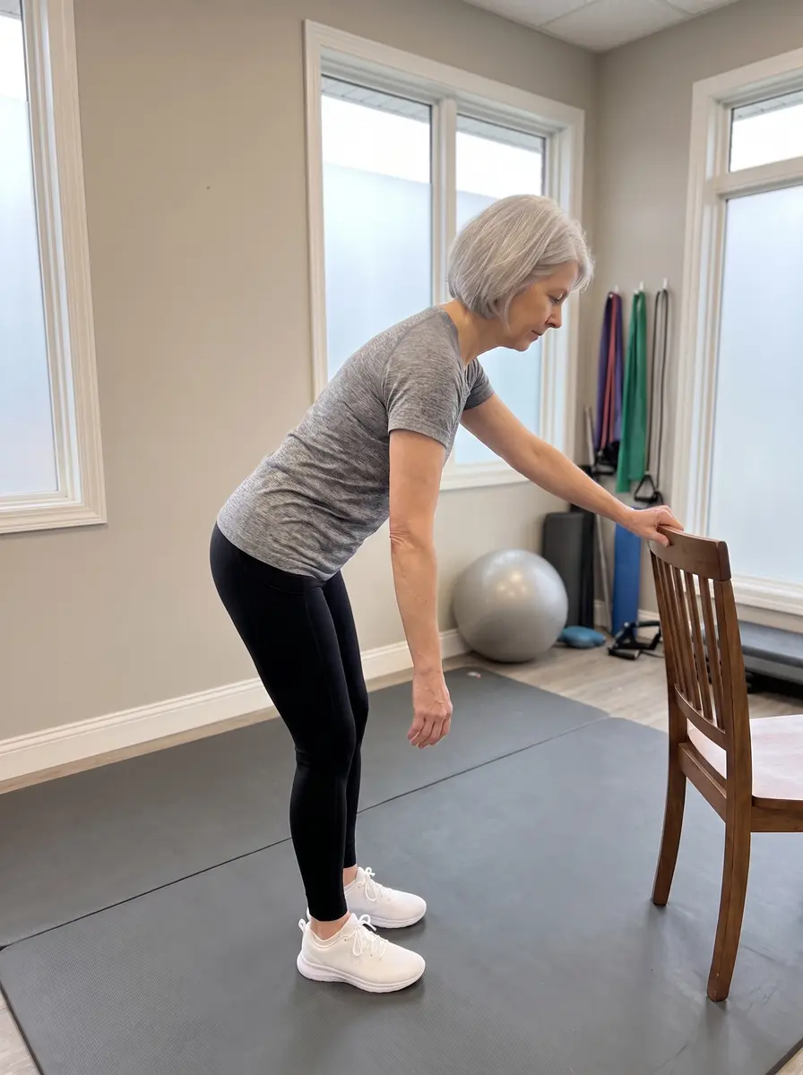
PendulumJoint & muscle
Lean on a chair, let the sore arm hang heavy, and swing it in small relaxed circles. One minute each direction.
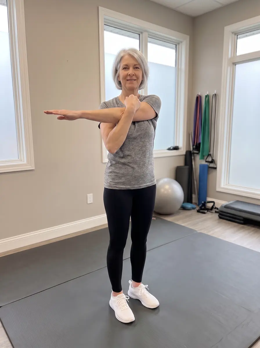
Cross-Body StretchJoint & muscle
Draw one arm across the chest and hug above the elbow. Hold 20–30 seconds; 3 times each side.
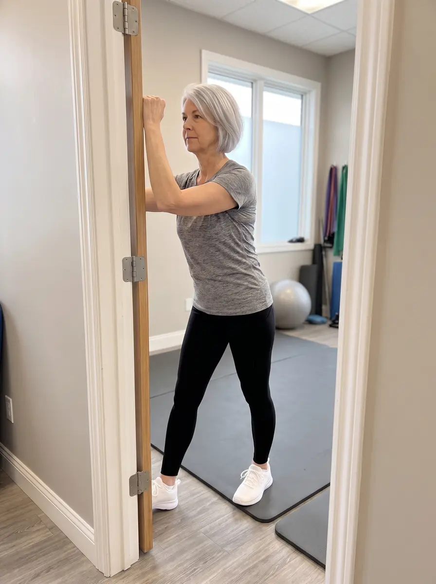
Doorway StretchJoint & muscle
Forearm on the frame, elbow at shoulder height, step gently through. Hold 20–30 seconds; 3 times.
🦾 Elbow & forearm
Tennis elbow is a tendon-loading problem, not simple inflammation — slow, heavy lowering is what rebuilds it. But burning pain with tingling that runs down FROM the neck is a nerve problem: treat the neck (see the nerve sliders above), not the elbow.
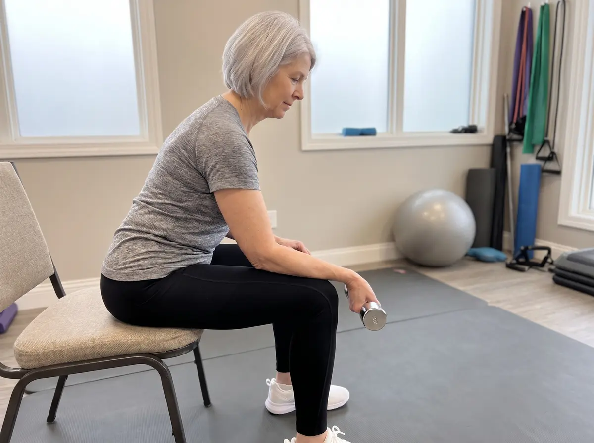
Eccentric Wrist LoweringJoint & muscle
Forearm supported palm-down, small weight in hand. Lower the wrist slowly over 3–5 seconds; use the other hand to help lift it back up. 3 sets of 10–15, daily — mild ache during is acceptable, sharp pain is not.
🦵 Hip
Deep buttock pain with tingling down the leg behaves like a NERVE problem — use the sciatic slider in the low-back group. The ache of hip arthritis lives in the groin or outer hip and loves strengthening.
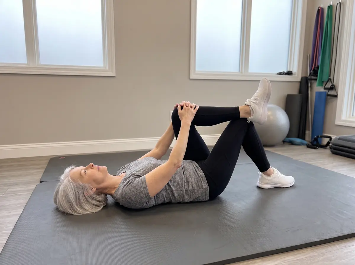
Figure-4 StretchJoint & muscle
Cross one ankle over the opposite knee and pull that thigh toward the chest until the buttock stretches. Hold 20–30 seconds; 3 times each side.
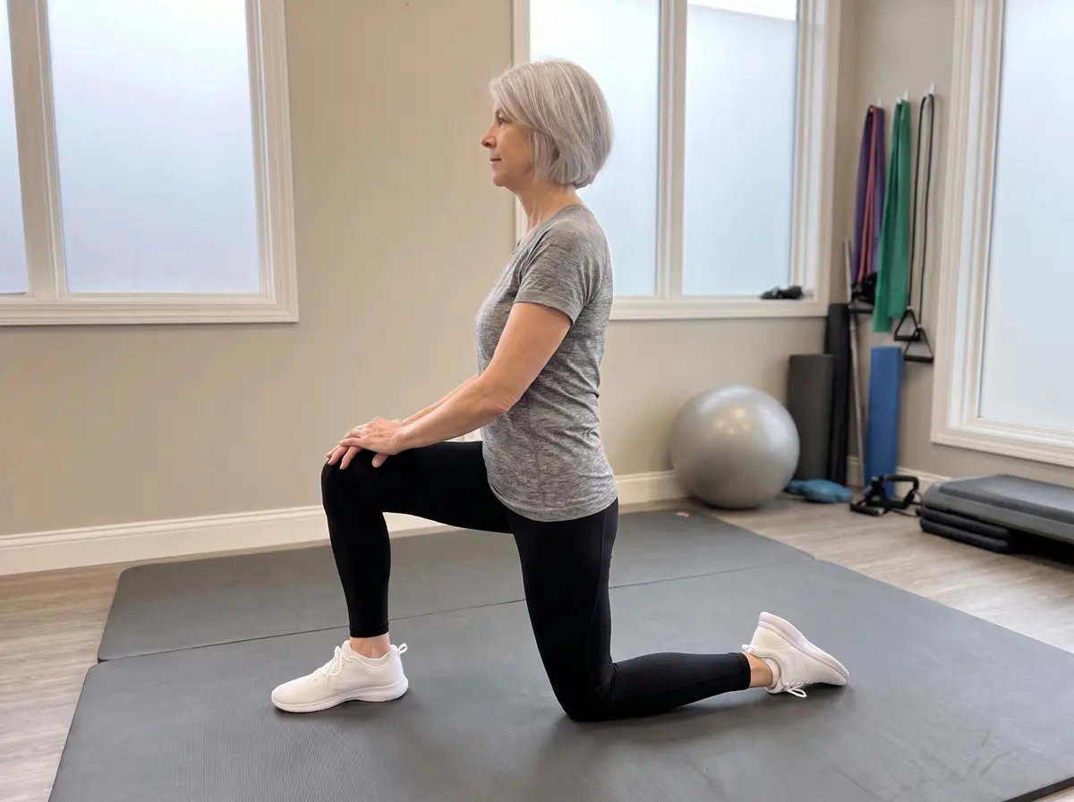
Kneeling Hip-Flexor StretchJoint & muscle
Half-kneeling, tuck the tailbone and shift the hips forward until the front of the hip stretches. Stay tall. Hold 20–30 seconds; 3 times each side.
ClosedOpen
▶ Watch the movement
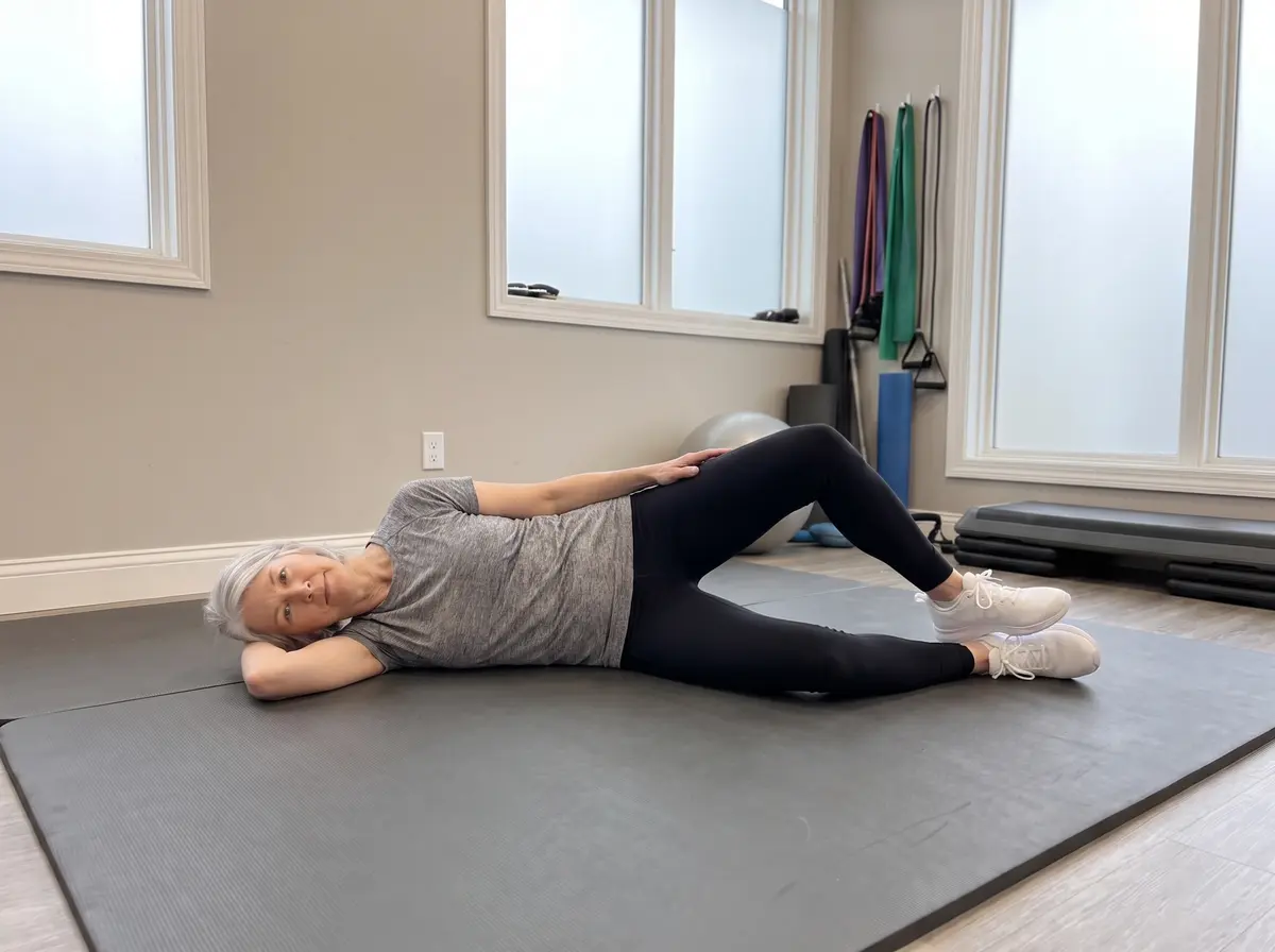
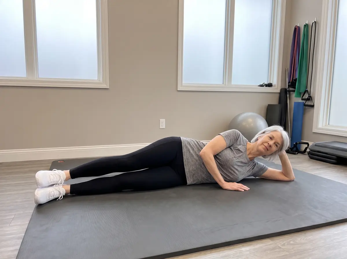
ClamshellJoint & muscle
Side-lying with knees bent and feet together, lift the top knee like a clamshell without rolling the pelvis back. 10–15 per side.
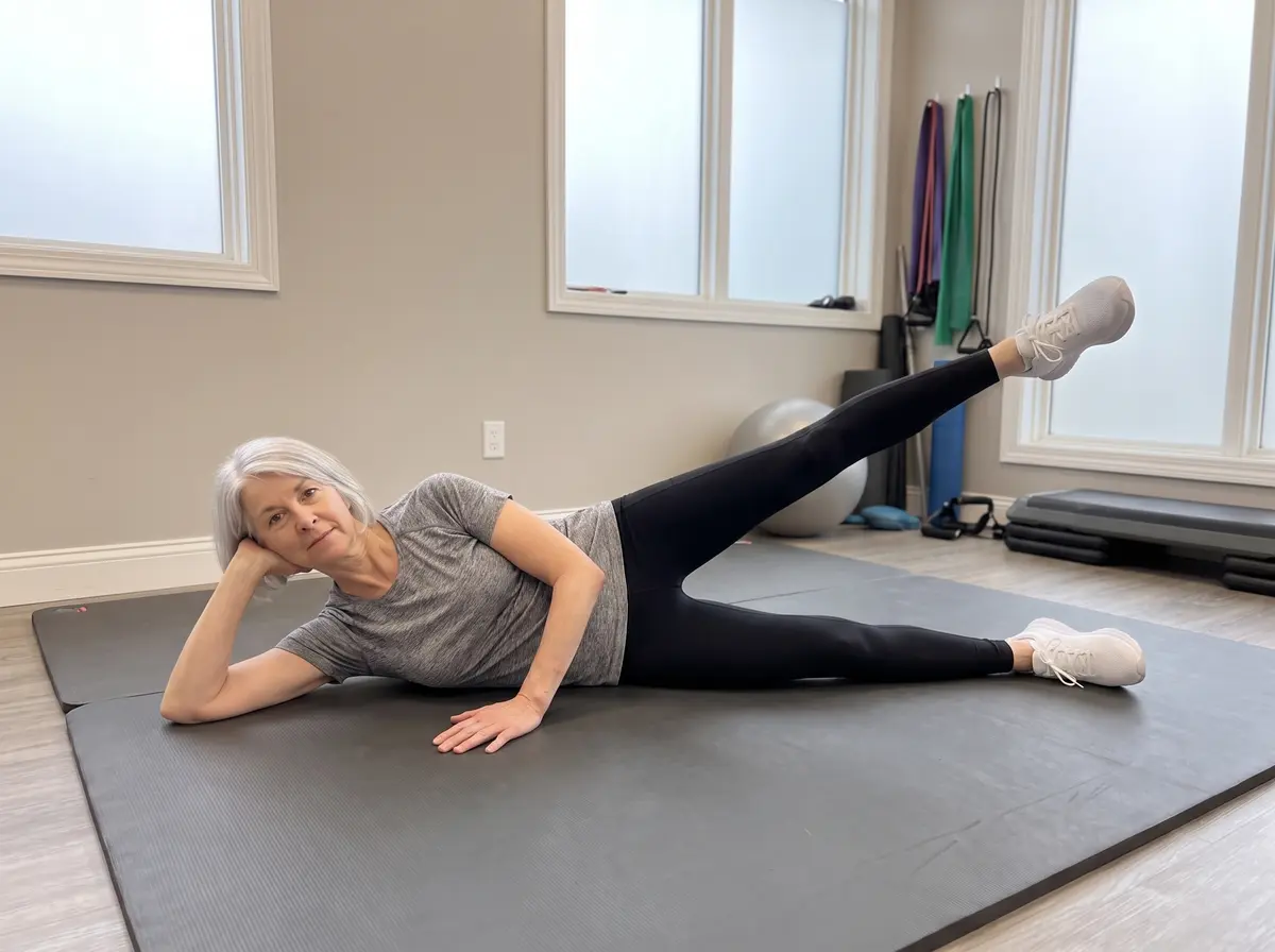
StartLift
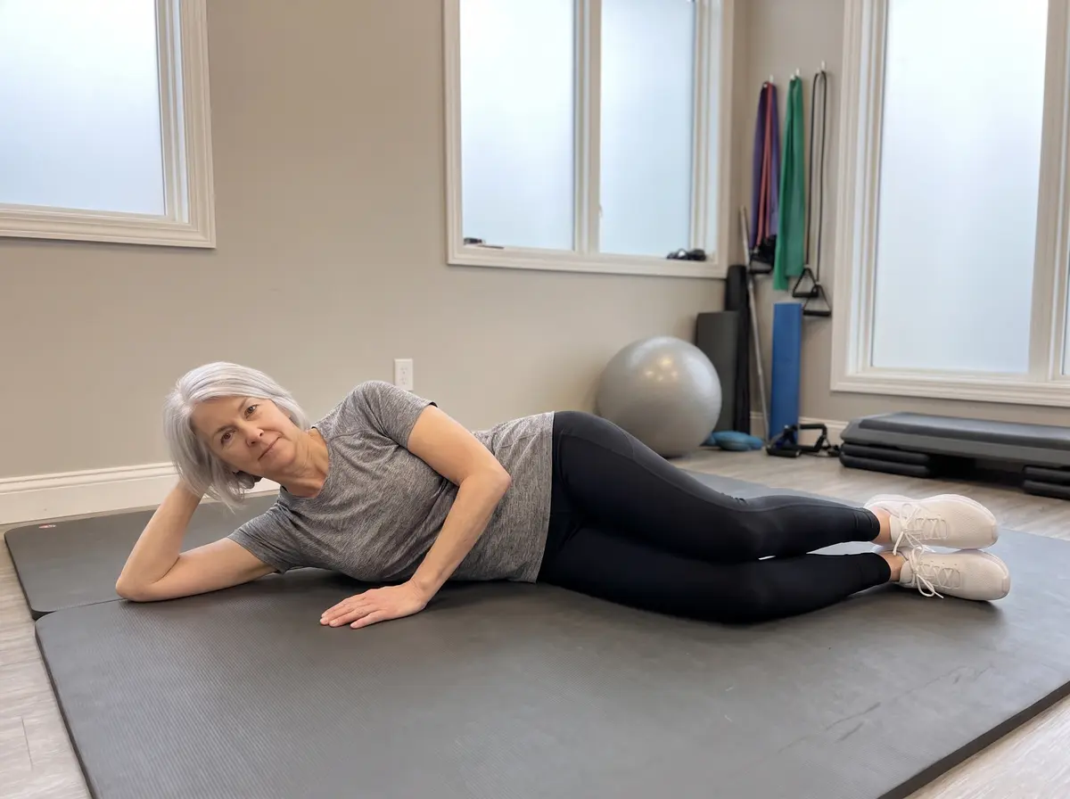
Side-Lying Leg RaiseJoint & muscle
Body in one straight line, lift the top leg to about 45° with toes forward, lower slowly. 10–15 per side.
🦿 Knee
The strongest medicine for an arthritic knee is the muscle above it: a strong quadriceps and strong hips absorb the force the worn cartilage no longer can.
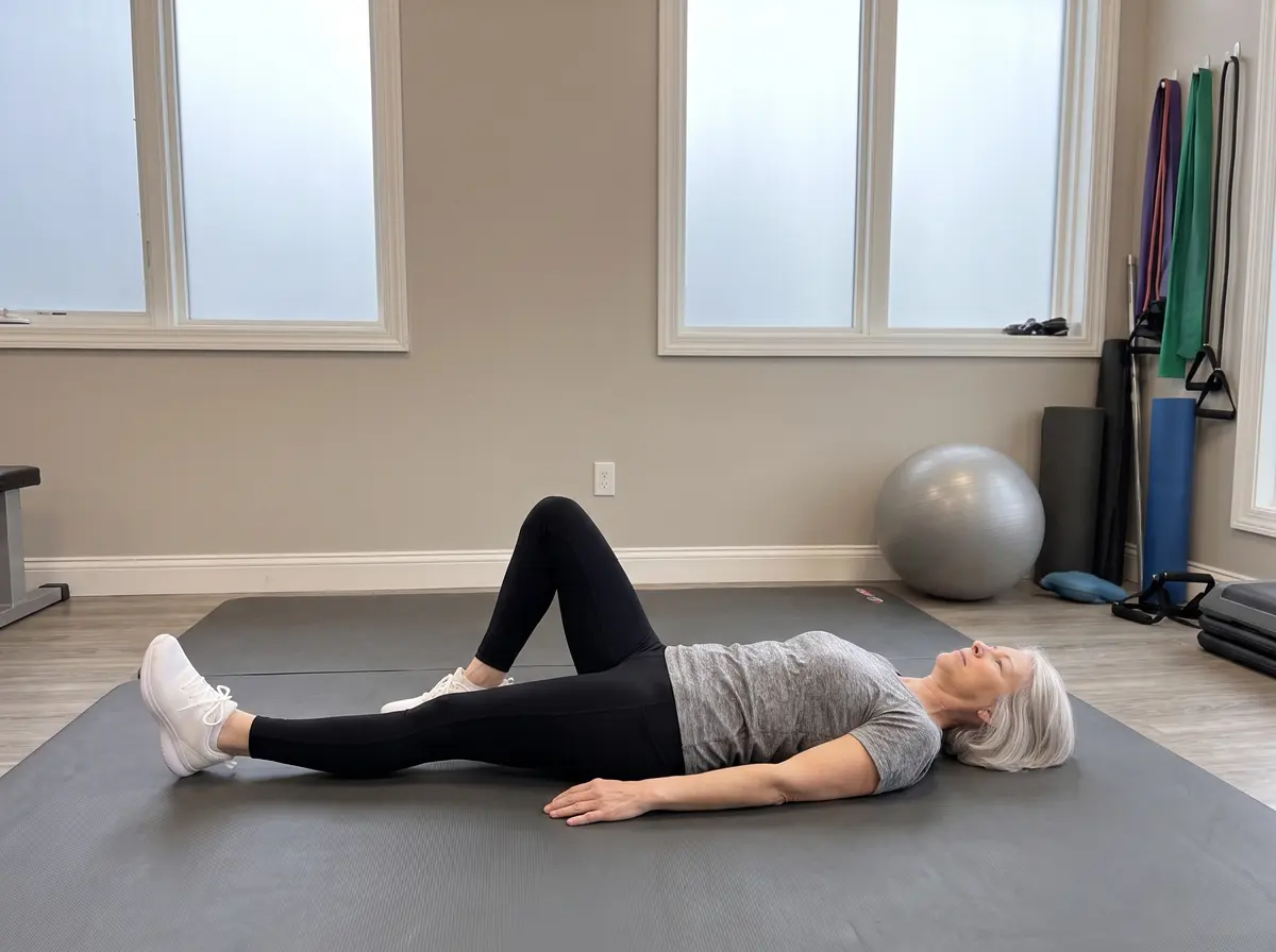
StartLift
▶ Watch the movement
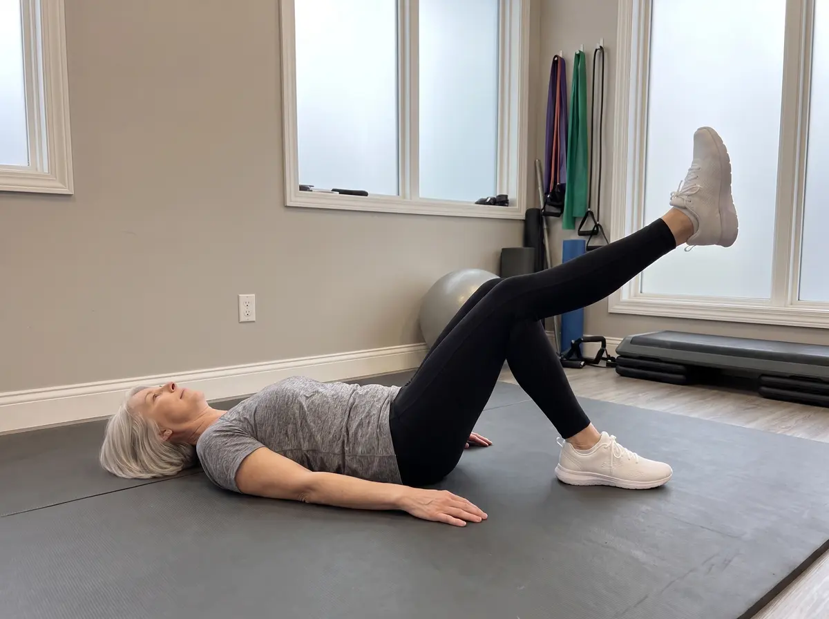
Straight-Leg RaiseJoint & muscle
One knee bent, the other leg straight. Tighten the thigh, lift the straight leg to the height of the bent knee, lower slowly. 10 per side.
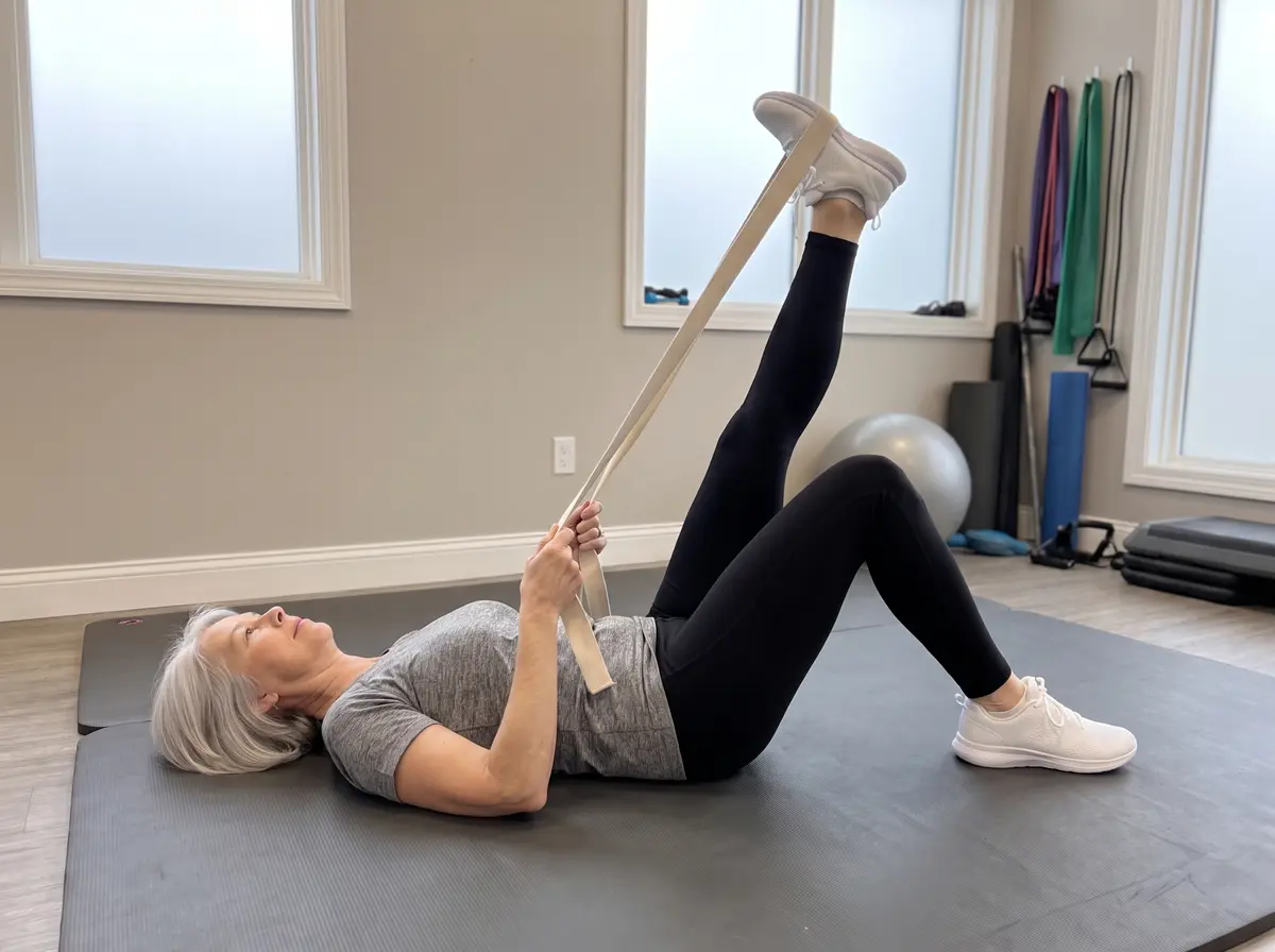
Hamstring Stretch with StrapJoint & muscle
Loop a strap around the foot and raise the straight leg until the back of the thigh stretches. Hold 20–30 seconds; 3 times each side. If this brings tingling or electric pain, your “tight hamstring” may actually be an irritated sciatic nerve — switch to the sciatic slider instead.
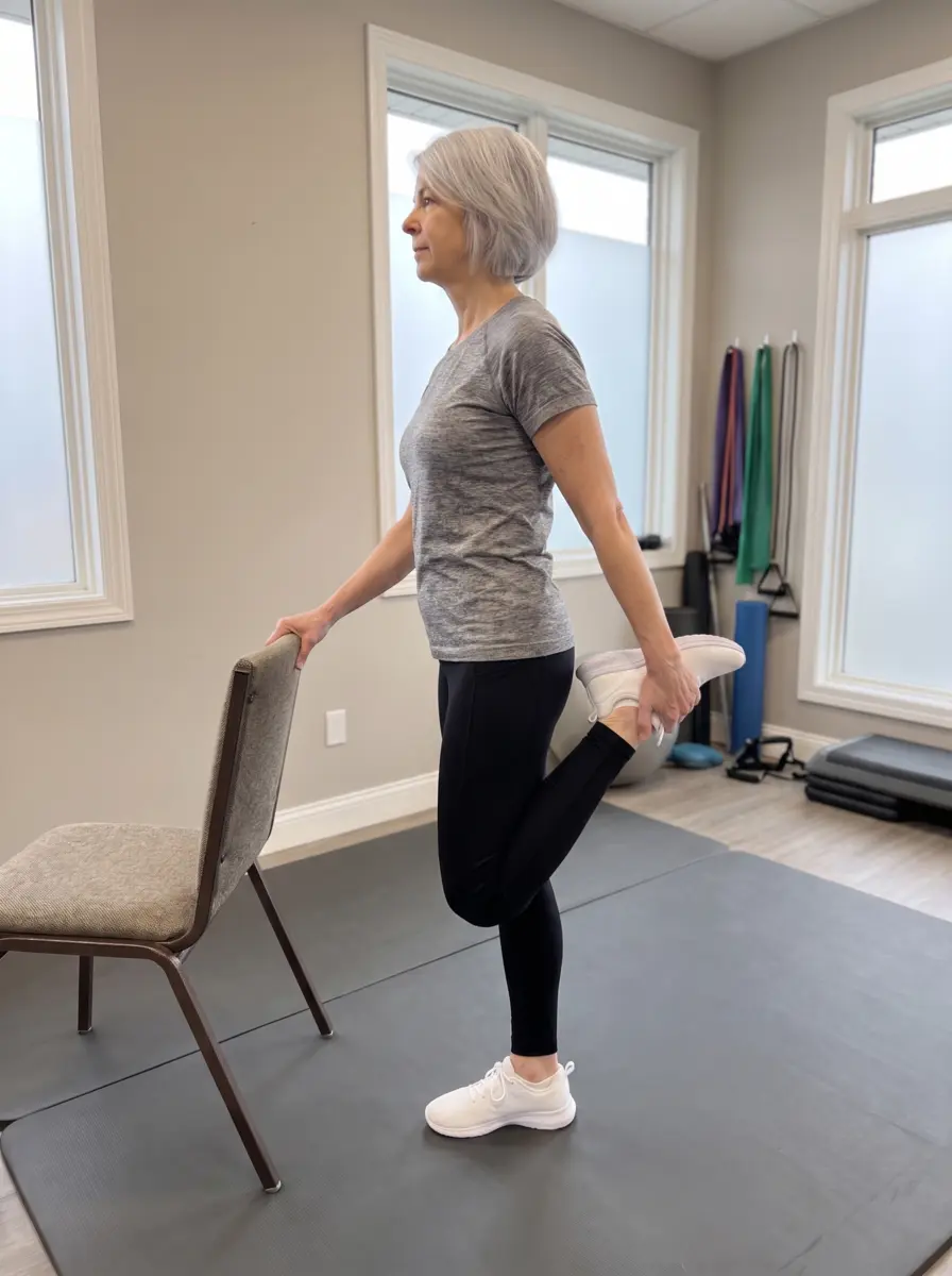
Standing Quad StretchJoint & muscle
Holding a chair, bring one heel toward the buttock, knee pointing down, standing tall. Hold 20–30 seconds; 3 times each side.
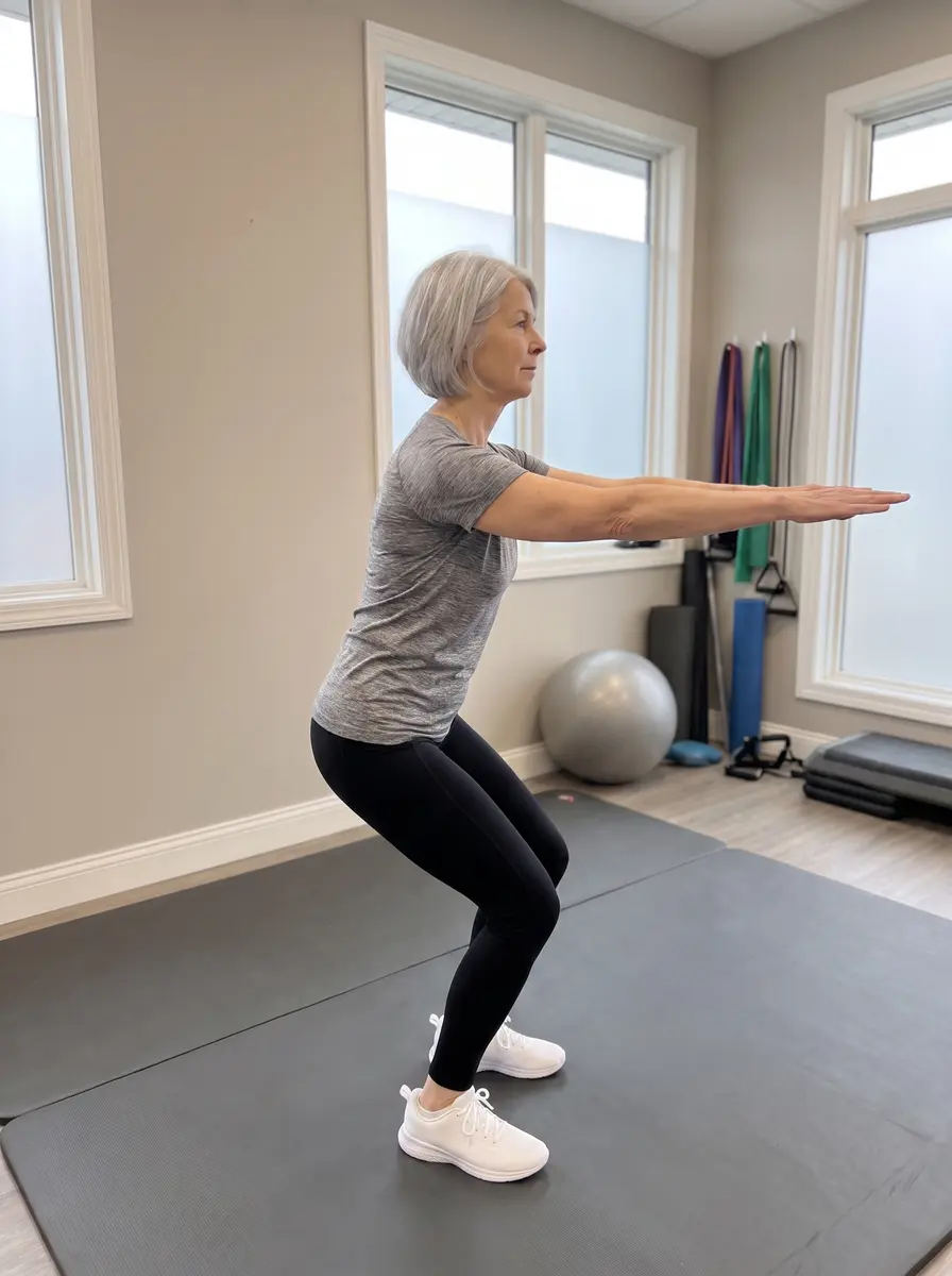
Mini SquatJoint & muscle
Feet shoulder-width, hips back, bend the knees about 45°, then stand tall. Knees track over the toes. 10–15 repetitions.
🦶 Foot & ankle
Heel and arch pain from plantar fasciitis responds to LOADING, not just stretching — the towel heel raise is the evidence-based centerpiece. Burning or numbness in the sole is more nerve-like; mention it, since it changes the plan.
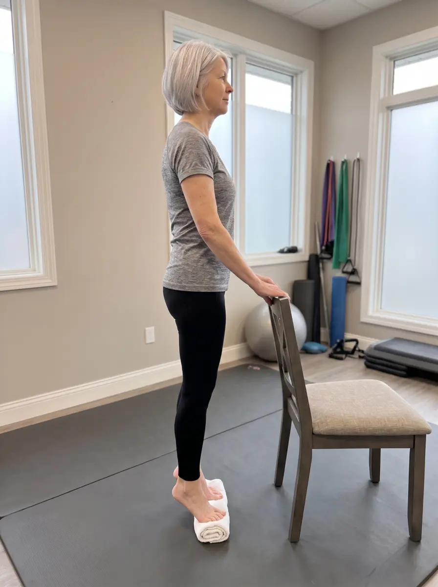
Heel Raise, Toes on TowelJoint & muscle
With a rolled towel under the toes (this tensions the arch), rise slowly onto the balls of the feet over 3 seconds, pause, lower over 3 seconds. 3 sets of 8–12, every other day — the proven plantar-fascia rebuilder.
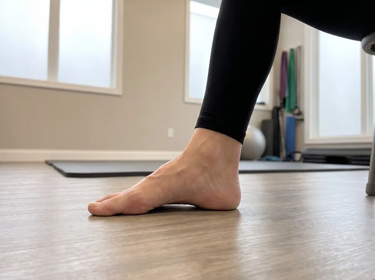
Short-Foot Arch LiftJoint & muscle
Seated, foot flat: dome the arch upward while the toes stay long and relaxed — as if shortening the foot. Hold 5 seconds; 10–15 reps per foot.
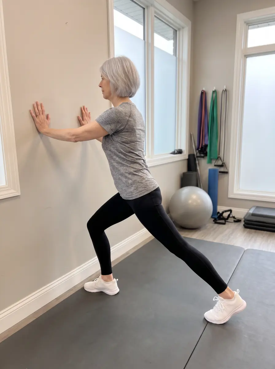
Wall Calf StretchJoint & muscle
Back leg straight, heel flat, lean in until the calf stretches. Hold 20–30 seconds; 3 times each side.
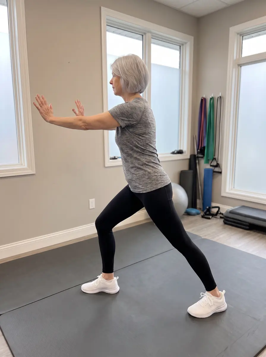
Soleus StretchJoint & muscle
Same stance with the back knee softly bent, heel down — the stretch drops toward the Achilles. Hold 20–30 seconds; 3 times each side.
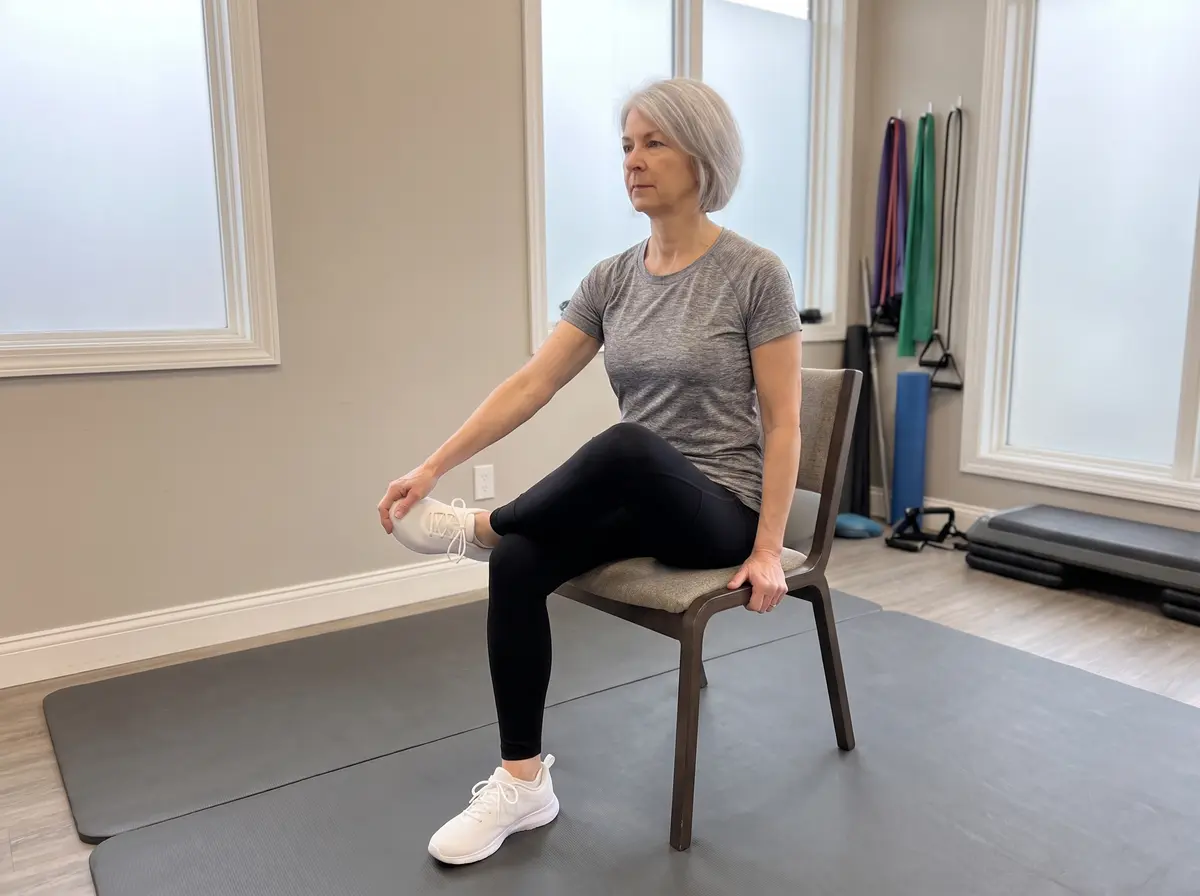
Plantar Fascia StretchJoint & muscle
Ankle crossed over the knee, pull the toes back until the arch stretches. Hold 20–30 seconds; 3 times — especially before the first steps of the morning.
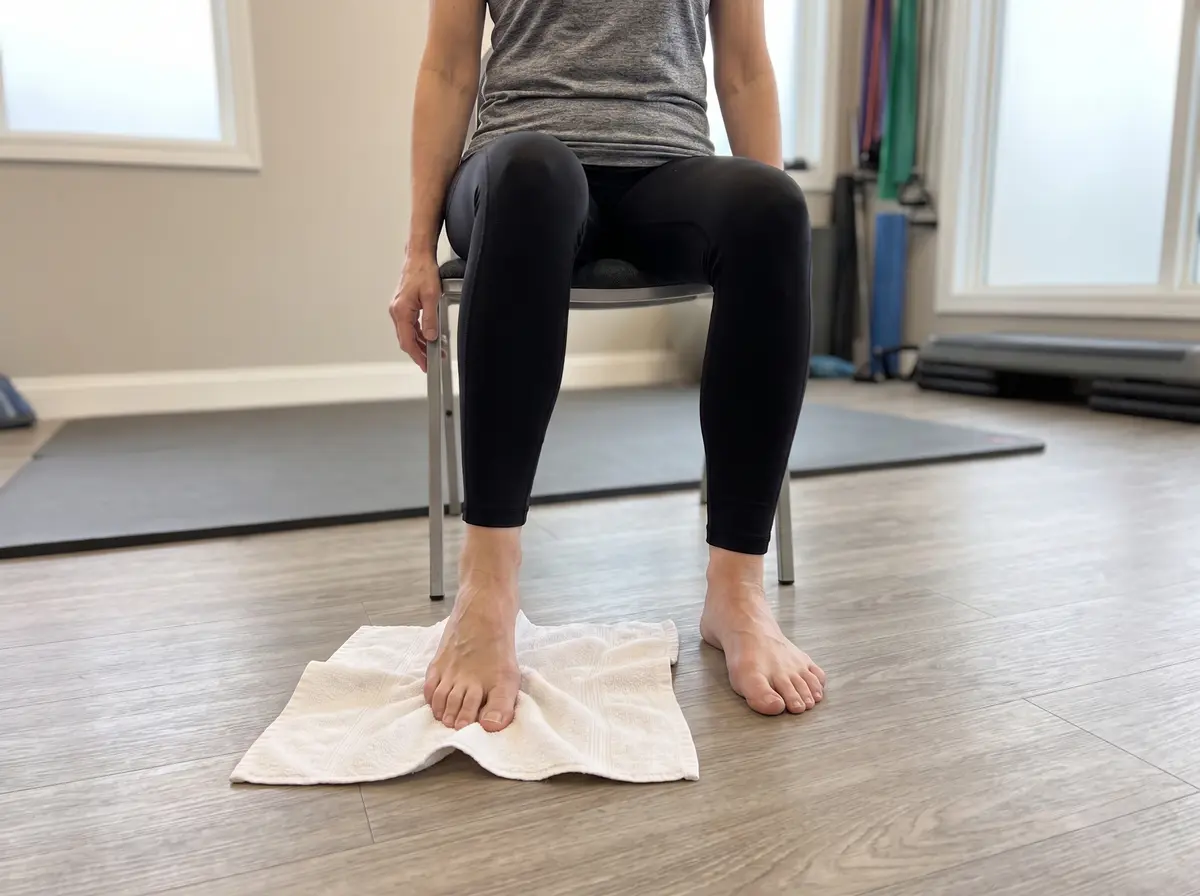
Towel ScrunchJoint & muscle
Barefoot, scrunch a towel toward you with the toes, re-flatten, repeat. About 2 minutes per foot.
StartRise
▶ Watch the movement
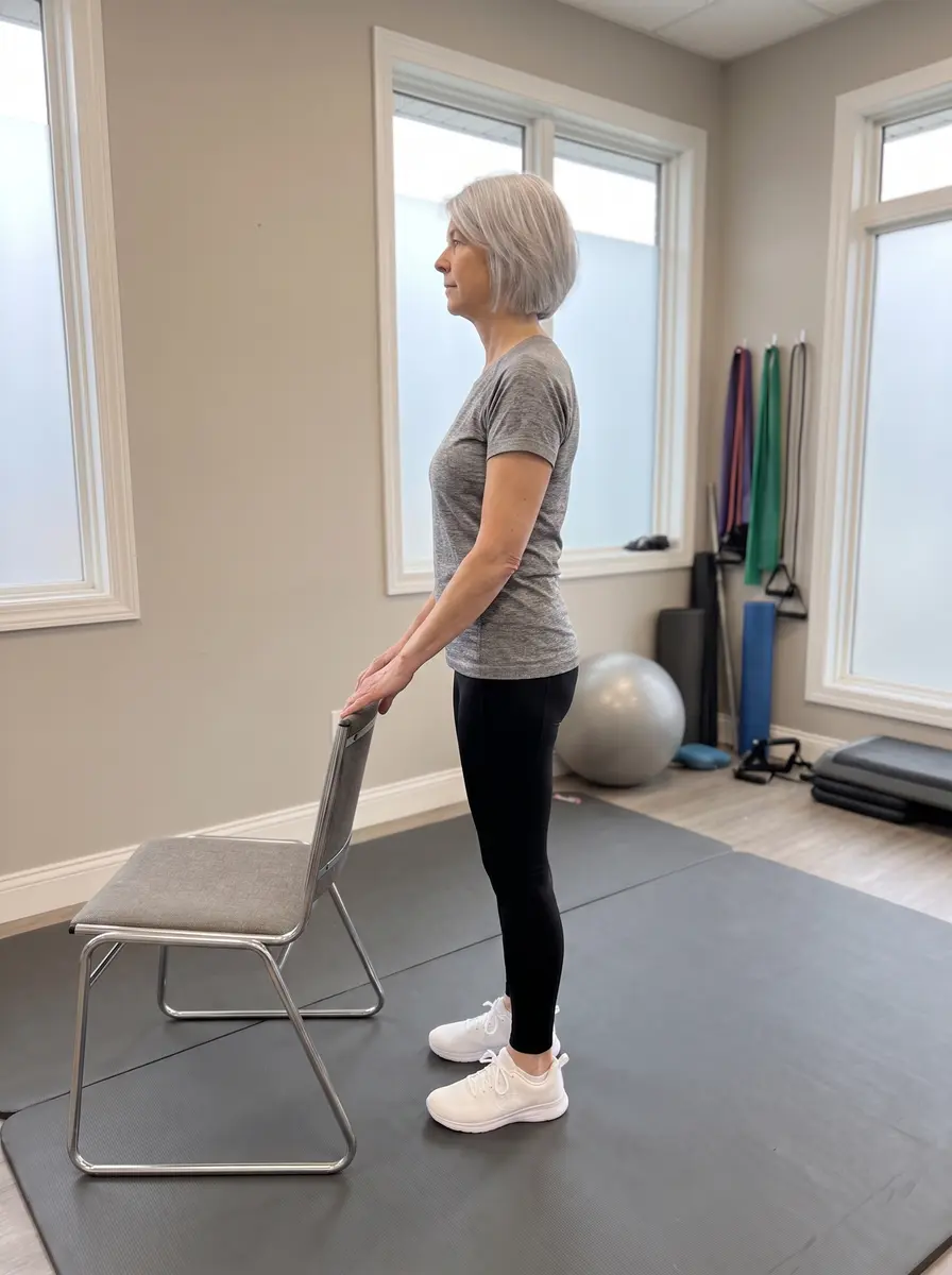
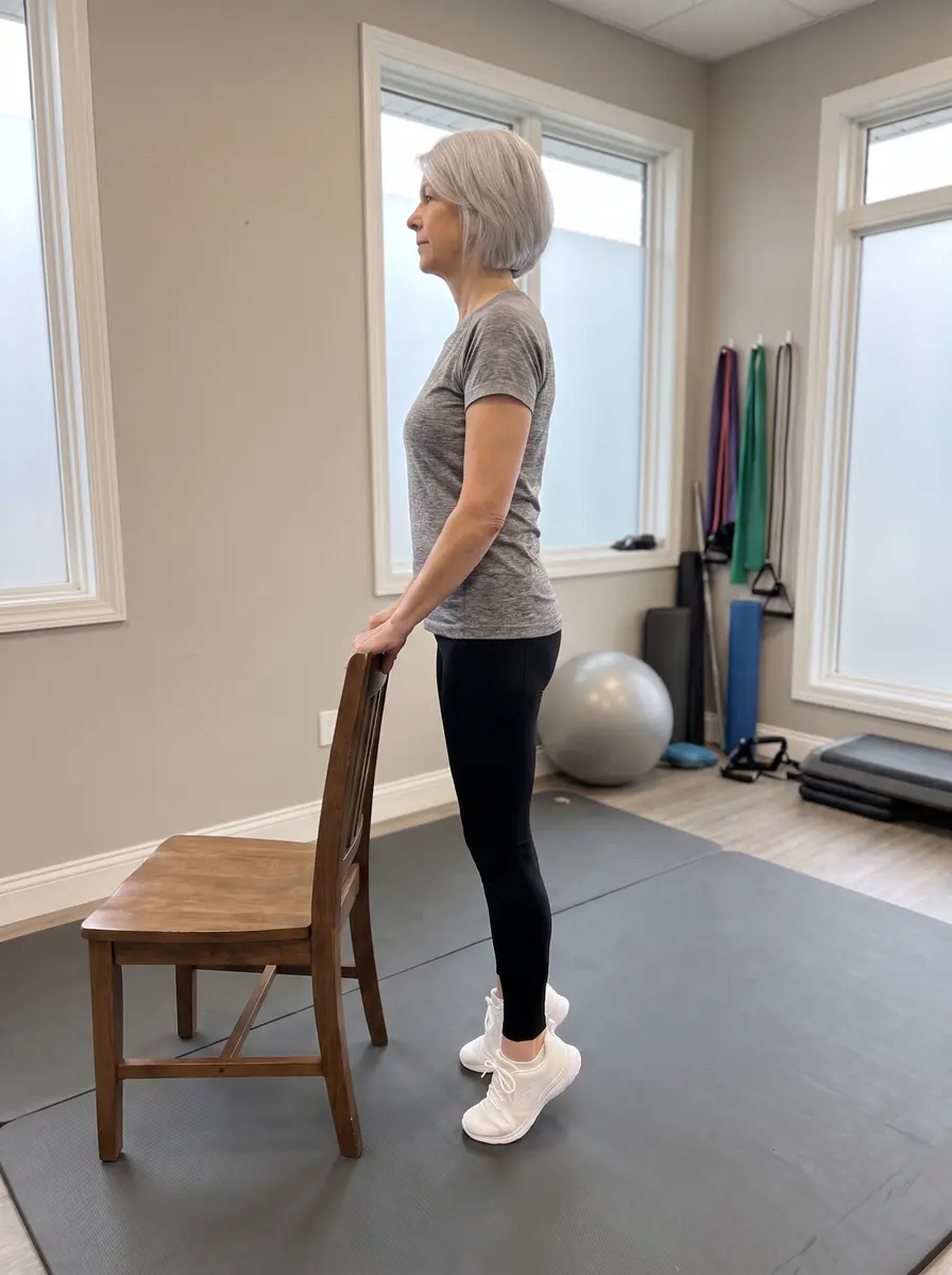
Heel RaisesJoint & muscle
Fingertips on a chair back, rise onto the balls of both feet, pause, lower with control. 10–15 repetitions.
Before you start: these exercises are general examples commonly used for each region and each pain type. Check with your clinician about which ones fit your specific condition — especially whether yours is a nerve target or a joint target — and stop any exercise that sharply increases your pain or sends it further down an arm or leg.
This page is educational material only and is not medical advice, a diagnosis, or a treatment recommendation. Illustrations are simplified artistic renderings, not exact anatomy; photos and videos are AI-generated demonstrations. Procedures, benefits, risks, and recovery vary from person to person — discuss your individual situation with your physician. In an emergency, call 911.