Medial branch radiofrequency ablation
Also called facet nerve ablation or medial branch neurotomy. When painful facet joints are confirmed with test blocks, gentle heat is used to quiet the tiny nerves that carry pain from those joints, often giving months of relief.
- Two test blocksUsually required first
- 6–12+ monthsTypical duration of relief
- RepeatableNerves regrow, so it can be repeated
- OutpatientHome the same day
What is a facet joint?
Each vertebra in your spine connects to the one above and below it in three places: the disc in front, and two small facet joints at the back, one on each side. Facet joints are about the size of a thumbnail. They are lined with cartilage, wrapped in a joint capsule, and they guide and limit how your spine bends and twists.
Like a knee or a finger joint, a facet joint can wear down with age (arthritis) or be strained by an injury. When that happens it can become a source of back or neck pain.
What facet pain typically feels like
- An ache on one or both sides of the spine, rather than pain shooting far down an arm or leg
- Often worse with leaning backward, twisting, or standing for long periods
- Stiffness first thing in the morning or after sitting
- Low back facet pain can spread into the buttock or upper thigh. Neck facet pain can spread to the shoulder blade or the back of the head.
No single sign on an exam or an MRI proves that a facet joint is the cause. That is why numbing injections are used to test it: if pain drops sharply after a joint or its nerves are numbed, that joint is likely part of the problem.
The medial branch nerves
Each facet joint gets its feeling from two tiny nerves called medial branches. They carry pain signals from the joint, and they also supply a few small back muscles. Importantly, they do not control the muscles of your arms or legs, and they do not carry feeling from your skin there. That is why they can be treated without causing weakness or numbness in your limbs.
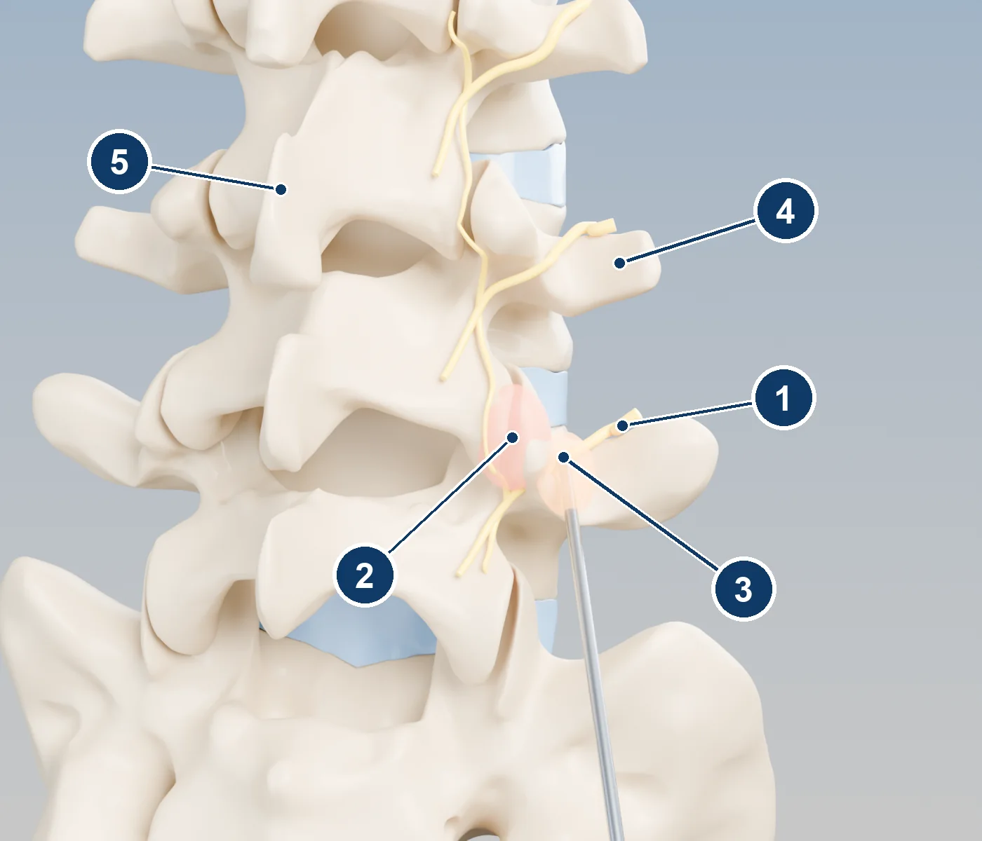
- 1Medial branch nerve running over the bone
- 2Facet joint, shown inflamed (shaded red)
- 3Radiofrequency probe tip and heating zone
- 4Transverse process, the bony "wing" on the side of the vertebra
- 5Spinous process
The usual pathway
Because no scan can prove that a facet joint is the pain source, treatment follows careful test steps.[4],[5]
At least 3 months of other care
Physical therapy, a home exercise program, and medicines first.
First medial branch block
A small amount of numbing medicine is placed on the medial branches. You keep a pain log for the next several hours.
Second, confirming block
Done on another day. Medicare in many regions requires at least 80% relief of your usual pain after each block before approving ablation. Other insurers use similar rules.
Radiofrequency ablation
If both blocks clearly helped, the same nerves are treated with heat for longer-lasting relief.
Repeat if needed
Nerves slowly regrow, so pain can return. Treatment can be repeated. Medicare generally covers a repeat if the first one gave at least 50% relief for at least 6 months.
How the ablation works
Using live X-ray, the physician places a thin, insulated needle-probe alongside each target nerve. The probe tip is heated with radiofrequency energy to about 80°C (176°F) for 60–90 seconds or more, making a small heat lesion that stops the nerve from carrying pain signals. Some systems use cooled or pulsed radiofrequency, but the principle is the same. Most treatments cover two to four nerves on one or both sides.
Procedure day
- Follow your eating and drinking instructions, and bring a driver.
- Do not stop blood thinners unless your prescribing doctor and procedure team tell you to.
- The skin is numbed. Some people are also given light sedation, though many regions limit sedation for this procedure.
- Before heating, a small test current checks that the probe is away from the nerves to your arms or legs. You may feel a tapping or buzzing in your back.
- Numbing medicine is given at each spot before heating. The procedure takes about 30–90 minutes.
Recovery
- Soreness, a burning or sunburn-like feeling, or muscle spasm near the treated area is common for 1–3 weeks. Ice and your usual pain relievers help.
- Relief usually builds over 2–4 weeks, so don’t judge the result in the first few days.
- Walk from the first day. Return to normal activity as soreness allows, and restart physical therapy when your team advises.
- Relief typically lasts 6 to 12 months or longer. As the nerves regrow, pain may return and the treatment can be repeated.
What the research shows
Neck facet pain vs sham (NEJM)
In 24 patients whose neck facet pain was confirmed with placebo-controlled blocks, pain took a median of 263 days to return after real ablation, compared with 8 days after a sham procedure.
Carefully selected low back patients
Among 106 patients selected by complete relief from two blocks, 53–58% had complete pain relief lasting at least 6 months, with a return to normal activities. Relief lasted about 15 months, and repeat treatments worked as well.
Large Dutch trial (MINT)
In 681 patients selected with a single block, adding ablation to an exercise program did not produce a clinically important extra benefit at 3 months. Specialists have noted that the looser selection and technique may explain the result.
International guideline
Lumbar medial branch ablation may benefit carefully selected patients. Stricter selection with two blocks improves results.
The honest summary: results depend heavily on choosing the right patients, which is why two test blocks come first. For people whose pain clearly comes from the facet joints, ablation can give months of relief. It is not a permanent cure, and it works best alongside exercise.
Risks
- Temporary increase in pain, or a burning, sunburn-like sensation over the treated area
- Bruising or bleeding
- Infection (rare)
- Small areas of skin numbness over the back
- Weakness of the small back muscles supplied by the treated nerves, usually without noticeable effect
- Rarely, injury to a nearby spinal nerve, causing leg or arm pain, numbness, or weakness
- Not getting relief, especially if another structure is also causing pain
If you have a pacemaker, defibrillator, spinal cord stimulator, or other implanted device, tell your team. Extra precautions are needed.
Call your care team right away if you have
- Fever of 100.4°F (38°C) or higher
- Redness, warmth, swelling, drainage, or bleeding at a treatment site
- New weakness or numbness in your arms or legs
- New loss of bladder or bowel control. Go to the emergency room.
Questions to bring
- How much relief did my blocks give, and was it enough?
- Which levels and sides will be treated?
- Will I have sedation, or just local numbing?
- When can I restart physical therapy?
- How will we know whether a repeat treatment makes sense?
References
- Lord SM, et al. Percutaneous radio-frequency neurotomy for chronic cervical zygapophyseal-joint pain. New England Journal of Medicine. 1996;335:1721-1726. doi.org/10.1056/NEJM199612053352302
- MacVicar J, et al. Lumbar medial branch radiofrequency neurotomy in New Zealand. Pain Medicine. 2013;14:639-645. doi.org/10.1111/pme.12000
- Juch JNS, et al. Effect of radiofrequency denervation on pain intensity among patients with chronic low back pain: the MINT randomized clinical trials. JAMA. 2017;318:68-81. doi.org/10.1001/jama.2017.7918
- Cohen SP, et al. Consensus practice guidelines on interventions for lumbar facet joint pain from a multispecialty, international working group. Regional Anesthesia & Pain Medicine. 2020;45:424-467. doi.org/10.1136/rapm-2019-101243
- Centers for Medicare & Medicaid Services. Local Coverage Determination L38801: Facet Joint Interventions for Pain Management (example regional Medicare policy). www.cms.gov/medicare-coverage-database/view/lcd.aspx?LCDId=38801
Related guides
About this page. This guide is general education written for patients. It does not replace advice from your own doctor, and results differ from person to person. Numbers quoted come from the published studies listed in the references; many large device studies are funded by the companies that make the devices, which we note where it applies.
Brand names that appear here are trademarks of their owners. This page is independent: it was not produced, paid for, or reviewed by any device manufacturer.
3D illustrations were rendered for this site from the open anatomy models of Z-Anatomy and BodyParts3D (© The Database Center for Life Science, CC BY-SA 2.1 JP; Z-Anatomy, CC BY-SA 4.0). Device hardware is a generic representation, not any specific product. The illustrations are shared under CC BY-SA 4.0.
Last reviewed: September 2026. If you have urgent symptoms, call your care team or 911.