💉 Epidural steroid injections · 5
Anti-inflammatory medicine delivered into the epidural space — the protective sleeve around the spinal nerves — to calm an inflamed nerve root. Different doorways, same destination.
Lumbar Epidural — Interlaminar
The classic approach for sciatica, entering between the laminae at the midline
Anti-inflammatory medicine placed into the epidural space of the low back through the midline window between two vertebrae.
Often used for: Sciatica from disc herniation or stenosis, often with back pain.

Full patient guide: Lumbar epidural steroid injectionFor sciatica: leg pain from an irritated nerve root in the low back. How it works, the visit step by step, risks, and what the research shows, with sources.
📚Epidurals reliably shorten severe sciatica flares; large reviews show the biggest benefit in the first weeks to months. Liu C et al. · BMJ 2023 ↗
1The anatomy
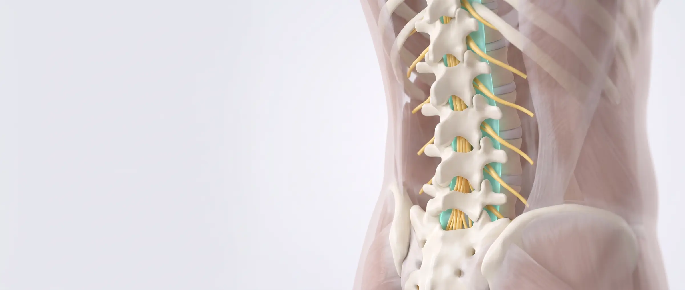
The epidural space (teal) surrounds and cushions the nerves of the low back.
2Where the pain starts
A herniated disc presses on a nerve root and inflames it — pain shoots down the leg.
3The procedure
Face down, skin numbed, a very thin needle guided by X-ray between the vertebrae.
4Relief delivered
Steroid and anesthetic bathe the inflamed root — outside the nerve sac — so it can settle.
What to expect: Home the same day; relief typically builds over 3–7 days.
Cervical Epidural — Interlaminar
The neck version, for nerve pain radiating into the shoulder or arm
The same medicine delivered into the epidural space of the neck, entering at its widest, safest level near the base.
Often used for: Pinched cervical nerves — arm pain, tingling, numbness from disc herniation or spondylosis.

Full patient guide: Cervical epidural steroid injectionFor arm pain from a pinched nerve in the neck. How it works, the visit step by step, risks, and what the research shows, with sources.
1The anatomy
Nerve roots exit between the neck vertebrae toward the shoulders and arms; the epidural space cushions the cord.
2Where the pain starts
A worn or herniated disc inflames an exiting root — pain travels along its path into the arm.
3The procedure
Face down with the forehead supported; a thin needle advanced under continuous X-ray at the base of the neck.
4Relief delivered
Medication spreads gently upward through the epidural space; the needle never touches the spinal cord.
What to expect: Mild soreness for a day or two; relief builds over the week.
Transforaminal Epidural (Nerve Root Injection)
Medicine delivered through the nerve's own doorway — the most targeted epidural
Instead of the midline, the needle approaches the small side window (foramen) where one specific nerve root exits — placing concentrated medicine exactly at the inflamed root.
Often used for: Sciatica or arm pain clearly traced to one nerve level; also used as a diagnostic test of which level is the culprit.
~15 minutesX-ray + contrastMost targeted option
1The nerve's doorway
Each nerve root leaves the spine through its own small window between the vertebrae — the foramen.
2A crowded doorway
A disc bulge or bone spur can narrow the foramen and pinch the root right in the doorway.
3The procedure
From an oblique angle, a thin needle rests at the outer opening of the foramen; contrast dye confirms it outlines the nerve sleeve.
4Relief delivered
Medication flows inward along the root sleeve into the epidural space — full strength, exactly where the problem lives.
What to expect: Brief pressure or a familiar 'zing' down the limb can happen as contrast spreads — expected and useful. Relief builds over days.
Caudal Epidural
Entering from below through the tailbone's natural opening
The epidural space is reached through the sacral hiatus — a natural opening at the very base of the spine — letting a larger volume of medicine wash upward over many nerve roots.
Often used for: Multi-level stenosis, post-surgical backs where scar changes the anatomy, and when other windows are hard to enter safely.
~15 minutesX-ray guidedGood after prior surgery
1The anatomy
At the bottom of the sacrum sits a small natural opening between two bony horns — the sacral hiatus.
2The procedure
A thin needle slips through the hiatus at a shallow angle into the sacral canal — far below where the nerve sac ends.
3Relief delivered
A generous volume of medication spreads upward through the epidural space, bathing several roots at once.
What to expect: Pressure at the tailbone during injection is normal. Home the same day.
Epidural Lysis of Adhesions (Racz)
Frees nerves trapped in post-surgical scar tissue
A steerable catheter is guided into the epidural space to mechanically open scarred compartments and deliver medication directly to nerve roots that standard epidurals can no longer reach.
Often used for: Post-laminectomy pain, epidural scarring, failed standard epidurals.
~45 minutesSteerable catheterTargets scarred segments
1How it works
Frees nerves trapped in post-surgical scar tissue
2The result
Sometimes done as a series.
What to expect: Sometimes done as a series. Relief comes from freeing the root and bathing it in medication where scar had walled it off.
🦴 Facet & SI joint treatments · 5
For the small joints of the spine and the two large joints where it meets the pelvis — test first, then treat.
Lumbar Medial Branch Blocks → RFA
Test the joints, then switch off their pain nerves with precise heat
Each facet joint reports pain through two tiny medial branch nerves. Numbing them is the test; radiofrequency heat is the durable treatment.
Often used for: Facet arthritis pain — worse standing, arching, twisting.

Full patient guide: Medial branch radiofrequency ablationMonths of relief by quieting the nerves to painful facet joints. How it works, the visit step by step, risks, and what the research shows, with sources.
1The pain source
Arthritic facet joints ache through their tiny medial branch nerves.
2The test block
A drop of numbing medicine at each branch. Substantial relief for hours = joints confirmed.
3Radiofrequency ablation
A heated tip creates a millimeters-wide lesion alongside each confirmed nerve.
4Lasting relief
The joint's message no longer reaches the spine; nerves regrow slowly and it can be repeated.
What to expect: Up to a week of soreness after ablation, then steady relief over 2–4 weeks.
Cervical Medial Branch Blocks → RFA
The neck version — including headaches that start at the skull base
The identical two-step logic applied to the neck's facet joints.
Often used for: Facet-driven neck pain, stiffness, and cervicogenic headaches.
Full patient guide: Medial branch radiofrequency ablationMonths of relief by quieting the nerves to painful facet joints. How it works, the visit step by step, risks, and what the research shows, with sources.
1The pain source
Arthritic neck facets — commonly behind neck pain that's worse looking up or turning.
2The test block
Thin needles place numbing drops where each medial branch crosses its bony pillar.
3Radiofrequency ablation
One small, controlled heat lesion per confirmed nerve.
4Lasting relief
Typically 9–18 months; repeatable when the nerves regrow.
What to expect: A test block wearing off same-day is expected — it's a diagnostic.
Facet Joint Injection (Intra-articular)
Medicine placed directly inside the joint itself
A thin needle enters the facet joint capsule and delivers steroid directly inside — sometimes chosen for younger joints, inflammatory flares, or cysts.
Often used for: Painful facet joints, especially inflammatory flares; also confirms the joint as the source.
~15 minutesX-ray + contrastJoint-targeted

Full patient guide: Facet joint injectionTesting and treating the small joints at the back of the spine. How it works, the visit step by step, risks, and what the research shows, with sources.
1Into the joint
Under X-ray, the needle slips into the small joint space; a crescent of contrast confirms it's inside the capsule.
2The result
Relief from the anesthetic within hours (diagnostic), steroid effect over the week.
What to expect: Relief from the anesthetic within hours (diagnostic), steroid effect over the week.
Sacroiliac Joint Injection
Where the spine meets the pelvis
Image-guided anti-inflammatory injection into the SI joint.
Often used for: One-sided low-back/buttock pain — standing from a chair, stairs, rolling in bed.
1The anatomy
Two strong, ligament-wrapped joints transfer your upper-body weight into the legs.
2Where the pain starts
An inflamed SI joint causes one-sided buttock pain, sometimes to the thigh.
3The procedure
A thin needle enters the lower joint; contrast confirms placement.
4Relief delivered
Medication fills the joint and calms the inflammation.
What to expect: Anesthetic may give hours of early relief; steroid builds over the week.
SI Joint Radiofrequency (Lateral Branch RFA)
For SI pain that returns after successful injections
The SI joint's small lateral branch nerves along the sacrum's edge are quieted with a row of precise heat lesions — the longer-lasting follow-up when injections work but wear off.
Often used for: Recurrent, injection-confirmed SI joint pain.
~30–45 minutesX-ray guidedRelief often 9–12+ months
1A row of quiet switches
Small cannulae line the sacral edge where the joint's tiny branches cross, each making one controlled lesion.
2The result
Soreness up to a week, then durable relief for many patients.
What to expect: Soreness up to a week, then durable relief for many patients.
🔥 Targeted nerve ablations · 3
When a specific small nerve is proven to carry the pain, precise heat or cold can silence it for months to years — while everything around it is protected.
Genicular Nerve Block → RFA (Knee)
Quieting the knee's pain wiring — without touching the joint
Three small genicular nerves carry pain from the knee capsule. After a numbing test confirms them, radiofrequency heat quiets each one at its bony landmark.
Often used for: Painful knee arthritis when surgery isn't wanted or isn't an option — and persistent pain after knee replacement.
Two visitsX-ray or ultrasoundWalk the same day
1The knee's pain wiring
Three small nerves curve around the knee — above-inner, above-outer, below-inner — carrying its pain signals.
2The worn joint
Arthritic cartilage and bone spurs make the joint ache; its little nerves report every step.
3Radiofrequency ablation
Three slender cannulae — one per nerve — each create a small, precise heat lesion.
4Walking comfortably
With the pain wiring quiet, the same knee carries you further; relief often lasts 6–12+ months.
What to expect: The knee itself is unchanged — this treats the pain, so keep strengthening (see the knee exercise program).
Basivertebral Nerve Ablation (Intracept)
For 'vertebrogenic' back pain that lives inside the bone itself
Some chronic low-back pain comes from the endplates of a worn disc's neighboring bone, reported by a tiny nerve INSIDE the vertebra. A one-time probe quiets that nerve at its trunk.
Often used for: Chronic midline low-back pain with characteristic endplate changes on MRI (Modic changes), present for 6+ months despite conservative care.
One sessionImplant-freeDurable in trials (5+ yrs)

Full patient guide: Basivertebral nerve ablationA one-time, implant-free procedure for chronic central low back pain with Modic changes on MRI. How it works, the visit step by step, risks, and what the research shows, with sources.
1A nerve inside the bone
The basivertebral nerve branches like a small tree inside each vertebra, reporting wear at the endplates.
2The ablation
Through the pedicle, a curved probe reaches the nerve's trunk and quiets it with contained heat — nothing is left behind.
What to expect: Improvement typically consolidates over 6–12 weeks; trial data show multi-year durability.
Cryoneurolysis (Nerve Freezing)
A tiny ice ball pauses a painful nerve — it regrows over months
A probe forms a small precise ice ball around a painful peripheral nerve, pausing its signal for weeks to months while it slowly regrows — a gentle, repeatable option.
Often used for: Focal nerve pain: around the knee, occipital region, intercostal/post-thoracotomy pain, some neuromas.
Office-basedUltrasound guidedReversible by design
1The freeze
A frosted micro-ice-ball forms around the nerve, pausing conduction without cutting anything.
2The result
Numbness in that nerve's small territory is expected and temporary.
What to expect: Numbness in that nerve's small territory is expected and temporary.
🕸️ Sympathetic & visceral blocks · 5
The sympathetic chains are the body's 'automatic' nerves — circulation, sweating, visceral pain. Blocking the right junction box can switch off pain that ordinary nerves don't explain.
Stellate Ganglion Block
The neck's sympathetic junction box — for CRPS and nerve pain of the arm and face
Local anesthetic placed beside the stellate ganglion — the sympathetic relay at the base of the neck — resetting overactive circuits to the arm, face, and heart region.
Often used for: Complex regional pain syndrome of the arm, some facial and post-herpetic pain, circulation disorders; being studied for PTSD.
~15 minutesUltrasound/X-rayEffects can outlast the drug
1The junction box
A chain of small ganglia runs in front of the spine; the stellate sits at the neck's base, wired to the arm and face.
2The overactive circuit
In CRPS the automatic wiring misfires — the hand burns, swells, changes color and temperature.
3The block
A thin needle from the front of the neck bathes the ganglion in anesthetic — vessels and airway kept safely aside.
4The reset
The arm warms and calms as the circuit resets. A droopy eyelid for a few hours means the block worked.
What to expect: Temporary hoarseness or eyelid droop = expected signs of success. Blocks are often given as a series while therapy retrains the limb.
Lumbar Sympathetic Block
The same reset for the leg
Anesthetic placed along the sympathetic chain on the front-side of the lumbar spine — the relay for the leg's automatic circulation and pain amplification.
Often used for: CRPS of the leg, some vascular and post-herpetic pain, phantom limb pain.
~20 minutesX-ray guidedOften a series
1The chain
Beads of sympathetic ganglia run along the front of the lumbar vertebrae, wired to the leg.
2The result
A warm, flushed leg right after = the block is working.
What to expect: A warm, flushed leg right after = the block is working.
Celiac Plexus Block / Neurolysis
For deep upper-abdominal pain — especially cancer pain
The celiac plexus — the nerve web in front of the aorta serving the upper abdominal organs — is bathed in anesthetic (block) or, for cancer pain, longer-acting agents (neurolysis).
Often used for: Pancreatic and other upper-abdominal cancer pain; select chronic pancreatitis.
~30 minutesImage guidedCan cut opioid needs
1The web
A delicate nerve web wraps the aorta below the diaphragm, carrying pain from stomach, pancreas, liver.
2The block
Two thin needles from the back meet the web from both sides and envelop it in medication.
What to expect: Temporary blood-pressure dip or looser stools can follow — monitored. Many patients need less opioid after.
Ganglion Impar Block
The single midline junction for tailbone and pelvic-floor pain
A solitary ganglion in front of the tailbone serves the deep pelvis. One drop of medicine there can quiet coccyx and perineal pain.
Often used for: Coccydynia, perineal/rectal pain syndromes.
~10 minutesX-ray guidedSmall but mighty
1One small target
The ganglion impar nestles in front of the last coccyx segment; a fine needle reaches it from below.
2The result
Often combined with a tailbone cushion + pelvic-floor therapy plan.
What to expect: Often combined with a tailbone cushion + pelvic-floor therapy plan.
Superior Hypogastric Plexus Block
Quiets the nerve relay station for the pelvic organs
Local anesthetic (sometimes followed by neurolysis) placed at the nerve web in front of the lowest lumbar vertebra — the relay station carrying pain from bladder, uterus, prostate and rectum.
Often used for: Pelvic pain from cancer, endometriosis, or chronic pelvic pain syndromes.
~30 minutesX-ray or CT guidedOften repeated or made longer-lasting
1How it works
Quiets the nerve relay station for the pelvic organs
2The result
Home the same day.
What to expect: Home the same day. A diagnostic block that helps strongly may be followed by a longer-lasting version.
⚡ Neuromodulation · 5
Instead of blocking or burning, these therapies talk to the nervous system in its own language — electricity — and are always test-driven first.
Spinal Cord Stimulation
A pacemaker for pain — trialed for a week before any implant
Gentle electrical pulses delivered over the spinal cord mask pain signals before they reach the brain. A no-incision trial week always comes first.
Often used for: Persistent nerve pain after back surgery, painful neuropathy, CRPS.

Full patient guide: Spinal cord stimulator trialA 5-day test of stimulation over the spinal cord for nerve-type back and leg pain. How it works, the visit step by step, risks, and what the research shows, with sources.
1How pain travels
Chronic nerve pain climbs the spinal cord to the brain — soften it on the way up and the brain receives less.
2The trial — no incision
Temporary leads through a needle; a small external stimulator on a belt; you live your normal week.
3How it works
Gentle pulses scramble the ascending signal — soft tingling, or simply less pain.
4The implant
If the trial earns it (≥50% relief), a pacemaker-like battery goes under the skin. Remote controlled, removable.
What to expect: The trial IS the decision: you judge the benefit at home before anything permanent.
Peripheral Nerve Stimulation
The same idea, miniaturized to a single nerve
A fine lead placed alongside one painful peripheral nerve delivers gentle pulses that mask its signal — some systems are temporary (60 days), some permanent.
Often used for: Focal nerve pain: shoulder (suprascapular/axillary), knee, occipital, post-amputation, post-surgical.
Needle placementUltrasound guidedTemporary or implanted

Full patient guide: Peripheral nerve stimulationA thin lead beside one painful nerve, such as in the shoulder, knee, foot, or low back. How it works, the visit step by step, risks, and what the research shows, with sources.
1One painful nerve
A single nerve segment misfires; the rest of the limb is fine.
2The lead
Through a needle, a fine lead comes to rest alongside the nerve — no incision.
3Masking the signal
Gentle waves wash the nerve's red signal quiet; a wearable or small implant powers it.
What to expect: 60-day temporary systems can 'retrain' pain lastingly; implanted versions are for durable focal pain.
Intrathecal Pain Pump
Medicine delivered where it works — at a fraction of the oral dose
A small implanted pump feeds tiny, precise doses of medication directly into the spinal fluid — often achieving with milligrams what pills need grams to do, with fewer systemic side effects.
Often used for: Refractory cancer pain; select severe chronic pain and spasticity (baclofen).
Trial firstRefilled in officeProgrammable
1The target compartment
The fluid-filled intrathecal space bathes the spinal cord — medication here acts directly on pain pathways.
2Why so little goes so far
Oral dosing floods the whole body; intrathecal dosing whispers directly to the spine at ~1/100th the dose.
3The system
A slim pump under the abdominal skin, a fine catheter curving to the spine.
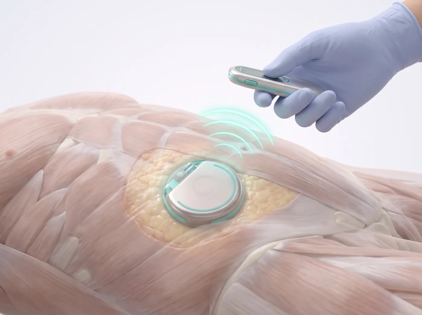
4Living with it
Refills by a simple office needle-stick every few months; dosing adjusted wirelessly.
What to expect: Always preceded by a trial dose that proves benefit first.
Dorsal Root Ganglion (DRG) Stimulation
Pinpoint neurostimulation for focal nerve pain
A cousin of spinal cord stimulation that parks a tiny electrode on the dorsal root ganglion — the nerve’s "junction box" — letting very focused areas like one foot, knee or the groin be treated precisely.
Often used for: CRPS of a foot or knee, groin pain after hernia surgery, other focal nerve injuries.
Trial first, like SCSImplanted if the trial worksVery focal coverage
1How it works
Pinpoint neurostimulation for focal nerve pain
2The result
You test-drive it for about a week first.
What to expect: You test-drive it for about a week first. DRG stimulation has landmark-trial evidence in focal CRPS.
Restorative Neurostimulation (Multifidus)
Retrains the spine’s deep stabilizing muscle
For chronic mechanical low back pain, an implanted device twitches the multifidus — the deep stabilizer that "switches off" in chronic back pain — twice a day, rebuilding its function over months.
Often used for: Chronic mechanical low back pain with poor muscle control, when therapy alone has stalled.
Implanted device30-minute sessions at homeBenefit builds over months
1How it works
Retrains the spine’s deep stabilizing muscle
2The result
Different philosophy from SCS: instead of masking pain signals it restores muscle control, so improvement accumulates gradually.
What to expect: Different philosophy from SCS: instead of masking pain signals it restores muscle control, so improvement accumulates gradually.
🏗️ Vertebral, disc & structural procedures · 8
When the problem is the architecture — a cracked vertebra, a canal grown too tight — these restore the structure through instruments the width of a pencil.
Kyphoplasty / Vertebroplasty
Stabilizing a painful spinal compression fracture from within
Through a pencil-width cannula, a small balloon re-expands the collapsed vertebra and medical cement stabilizes it — often turning severe movement-pain off within days.
Often used for: Painful osteoporotic (or tumor-related) vertebral compression fractures.
~30–45 minutesX-ray guidedOften rapid relief
1The fracture
A vertebral body collapses into a wedge — every movement grinds the crack, and posture tips forward.
2The balloon
Via the pedicle, a small balloon gently lifts the collapsed bone back toward its height.
3The cement
The space is filled with bone cement — the crack is knitted, the pain generator stabilized.
What to expect: Many patients feel dramatically better within 24–72 hours. Bone-health treatment matters just as much afterward.
MILD / Interspinous Spacer (Vertiflex)
More room for the nerves in lumbar stenosis — without open surgery
Two implant-light options for stenosis: MILD trims the thickened ligament through a tiny port; a Vertiflex spacer props open the space between spinous processes. Both aim to let you stand and walk longer.
Often used for: Lumbar spinal stenosis with classic 'shopping-cart' walking limitation.
OutpatientX-ray guidedMotion preserved
1Making room
Through a pencil-width tube, excess ligament is trimmed — the canal visibly opens and the nerves decompress.
2The result
Walking distance is the scoreboard — most gains show over the first weeks as the nerves calm.
What to expect: Walking distance is the scoreboard — most gains show over the first weeks as the nerves calm.
Provocation Discography
A diagnostic test that asks each disc directly: 'is it you?'
Contrast is injected into individual discs to see their internal structure and — crucially — whether pressurizing a specific disc reproduces YOUR familiar pain. Purely diagnostic, usually before surgical decisions.
Often used for: Selecting (or ruling out) discs before fusion or other structural decisions.
Diagnostic onlyX-ray + CTAnswers, not treatment
What to expect: A positive test = your exact pain reproduced at low pressure in one disc while neighbors stay quiet.
Sacroplasty
Cement reinforcement for a fractured sacrum
The sacral cousin of kyphoplasty: medical bone cement stabilizes insufficiency fractures of the sacrum — a common, underdiagnosed cause of severe sitting and walking pain in osteoporosis.
Often used for: Sacral insufficiency fractures, some sacral tumor-related fractures.
~45 minutesCT or X-ray guidedOften rapid relief
1How it works
Cement reinforcement for a fractured sacrum
2The result
Like kyphoplasty, pain relief can be dramatic within days as the fracture is internally splinted.
What to expect: Like kyphoplasty, pain relief can be dramatic within days as the fracture is internally splinted.
Interspinous Spacer
A small implant that holds the "standing room" open
For lumbar stenosis, a small H-shaped spacer placed between two spinous processes keeps the segment slightly flexed — preserving the canal space that opens when you sit — without removing bone.
Often used for: Neurogenic claudication from lumbar stenosis in patients who get relief by sitting or leaning forward.
~1 hour, outpatientSmall midline incisionReversible; preserves anatomy
1How it works
A small implant that holds the "standing room" open
2The result
Walk-in, walk-out decompression: standing and walking tolerance typically improve because the segment can no longer pinch shut in extension.
What to expect: Walk-in, walk-out decompression: standing and walking tolerance typically improve because the segment can no longer pinch shut in extension.
Minimally Invasive SI Joint Fusion
Stops the painful micro-motion of a failed SI joint
When a sacroiliac joint keeps failing diagnostic blocks-and-relief cycles, small titanium implants placed across the joint through a 2–3 cm incision stop its painful micro-motion permanently.
Often used for: Refractory SI joint dysfunction confirmed by repeated diagnostic blocks.
~1 hourImplants across the jointFor block-confirmed SI pain
1How it works
Stops the painful micro-motion of a failed SI joint
2The result
Reserved for well-confirmed SI pain.
What to expect: Reserved for well-confirmed SI pain. Most patients mobilize the same day with a walker during early healing.
Intradiscal Biacuplasty / IDET
Treats pain arising inside the disc itself
Two cooled radiofrequency probes placed in the back wall of a painful disc heat and quiet the small nerves that have grown into annular tears — treating discogenic pain at its source.
Often used for: Discogenic low back pain confirmed by exam, MRI pattern, and sometimes discography.
~1 hourX-ray guidedFor disc-origin pain
1How it works
Treats pain arising inside the disc itself
2The result
A niche but real option when the disc itself is the pain generator and surgery feels like too big a step.
What to expect: A niche but real option when the disc itself is the pain generator and surgery feels like too big a step.
Percutaneous Disc Decompression
Shrinks a contained herniation from the inside
A thin device removes a small channel of disc material from the center of a contained herniation, lowering the internal pressure so the bulge can retract off the nerve.
Often used for: Small contained herniations with leg pain, when ESIs help but keep wearing off.
~45 minutesOne thin deviceContained herniations only
1How it works
Shrinks a contained herniation from the inside
2The result
The disc stays intact; only a few percent of its core volume is removed — enough to decompress the nerve in well-chosen cases.
What to expect: The disc stays intact; only a few percent of its core volume is removed — enough to decompress the nerve in well-chosen cases.
💪 Muscle, tendon & small-nerve treatments · 10
The everyday workhorses of a pain clinic — quick, office-based, and often paired with the exercise programs in this site.
Trigger Point Injections
Releasing the knots that refer pain elsewhere
A fine needle (with or without medication) releases the taut, tender knots in muscle that ache locally and refer pain in predictable patterns.
Often used for: Myofascial pain of the neck, shoulders, and back — including 'headaches' that start in the trapezius.
Office-basedMinutesPairs with stretching
1The knot
A taut band with a dense, irritable knot — the trigger point.
2Referred pain
Trigger points send pain elsewhere along predictable maps — a shoulder knot can 'become' a temple headache.
3The release
A fine needle reaches the knot; a local twitch and a drop of medication let the band release.
4After
The muscle lies smooth again. Stretching and posture work keep it that way.
What to expect: Post-injection soreness for a day is normal; heat and gentle stretch help.
Botox for Chronic Migraine
31 tiny injections, every 12 weeks, fewer headache days
OnabotulinumtoxinA placed in a standardized pattern across forehead, temples, back of head, neck and shoulders reduces headache frequency in chronic migraine (15+ headache days/month).
Often used for: Chronic migraine; also used medically for severe spasticity elsewhere.
Every 12 weeksOffice-basedFDA-approved pattern
1The pattern
Hair-fine needles place micro-doses at the standardized sites; most patients rate it far easier than expected.
2The result
Benefit builds across the first two cycles — judge it at 24 weeks, not 2.
What to expect: Benefit builds across the first two cycles — judge it at 24 weeks, not 2.
Occipital Nerve Blocks
For headaches that climb the back of the skull
Anesthetic and steroid placed beside the greater/lesser occipital nerves at the skull base.
Often used for: Occipital neuralgia, cervicogenic headache, some migraine patterns.
MinutesLandmark/US guidedOften immediate
1The block
A drop of medication where the occipital nerves emerge — scalp numbness right after means the target was hit.
2The result
Relief within minutes (anesthetic phase) predicts steroid-phase benefit.
What to expect: Relief within minutes (anesthetic phase) predicts steroid-phase benefit.
Suprascapular Nerve Block
The shoulder's main sensory line
Medication placed at the scapular notch where the shoulder's chief sensory nerve passes — useful when the joint itself can't be settled.
Often used for: Frozen shoulder, cuff arthropathy, post-stroke shoulder pain.
MinutesUltrasound guidedRehab enabler
1The block
The nerve is bathed at its notch — often unlocking enough comfort to make therapy possible.
2The result
Best used as a bridge INTO the shoulder exercise program.
What to expect: Best used as a bridge INTO the shoulder exercise program.
Ilioinguinal / Iliohypogastric Block
For groin pain — often after hernia surgery
Ultrasound-guided medication placed in the muscle plane where these two nerves travel toward the groin.
Often used for: Post-herniorrhaphy groin pain, some chronic pelvic/groin neuralgias.
MinutesUltrasound guidedDiagnostic + therapeutic
1The plane
Medication spreads in the layer between abdominal muscles, enveloping both nerves.
2The result
If relief is real but temporary, cryo or PNS of the same nerves is the logical next step.
What to expect: If relief is real but temporary, cryo or PNS of the same nerves is the logical next step.
Pudendal Nerve Block
For deep pelvic pain with sitting
Image-guided medication beside the pudendal nerve at the ischial spine.
Often used for: Pudendal neuralgia — perineal burning worse with sitting, spared by standing.
~15 minutesImage guidedDiagnostic + therapeutic
1The block
The nerve is reached deep in the pelvis where it rounds the ischial spine.
2The result
Paired with pelvic-floor physical therapy for lasting change.
What to expect: Paired with pelvic-floor physical therapy for lasting change.
Carpal Tunnel Injection
Calming the median nerve in its tunnel
A small dose of steroid placed inside the carpal tunnel (beside, never into, the nerve).
Often used for: Carpal tunnel syndrome — night numbness, tingling thumb-to-ring fingers.
MinutesUS or landmarkSplint + glides after
1The tunnel
Medication spreads through the crowded tunnel, shrinking the swelling around the median nerve.
2The result
Night splinting and tendon-glide exercises extend the benefit.
What to expect: Night splinting and tendon-glide exercises extend the benefit.
Lateral Femoral Cutaneous Nerve Block
The burning-thigh nerve, calmed at the hip crease
The nerve that supplies the outer thigh skin gets pinched under the inguinal ligament (tight belts, pregnancy, weight change), causing meralgia paresthetica. Ultrasound guides medicine right around it.
Often used for: Meralgia paresthetica — burning, tingling outer thigh.
~15 minutesUltrasound guidedDiagnostic + therapeutic
1How it works
The burning-thigh nerve, calmed at the hip crease
2The result
Numbness of the outer thigh for a few hours confirms the diagnosis; steroid quiets it longer.
What to expect: Numbness of the outer thigh for a few hours confirms the diagnosis; steroid quiets it longer. Loose waistbands do the rest.
Intercostal Nerve Block / RFA
The rib nerves, numbed along their groove
Each rib carries a nerve in a groove along its lower edge. Blocks (and radiofrequency or cryo for longer relief) treat pain after rib fractures, chest surgery, or shingles of the chest wall.
Often used for: Rib fracture pain, post-thoracotomy pain, chest-wall neuralgia.
~20 minutesUltrasound or X-ray guidedCan be extended with RFA/cryo
1How it works
The rib nerves, numbed along their groove
2The result
Relief is often immediate.
What to expect: Relief is often immediate. Because the lung sits just deep to the nerve, imaging guidance is standard.
Piriformis Injection
Relaxes the deep muscle that irritates the sciatic nerve
The piriformis crosses directly over (or around) the sciatic nerve deep in the buttock. Image-guided injection of anesthetic and steroid — or botulinum toxin for stubborn cases — releases its grip.
Often used for: Piriformis syndrome: deep buttock pain with sciatica-like radiation.
~20 minutesUltrasound or X-ray guidedBotox for recurrences
1How it works
Relaxes the deep muscle that irritates the sciatic nerve
2The result
Pairs naturally with the stretching program — the injection opens a window; the exercises keep it open.
What to expect: Pairs naturally with the stretching program — the injection opens a window; the exercises keep it open.
🦵 Joint & bursa injections · 4
Image guidance turns a good injection into a precise one — medicine lands exactly in the joint or bursa, every time.
Hip Joint Injection
The deep joint that needs image guidance most
Steroid (or viscosupplement/PRP) delivered into the hip joint under X-ray or ultrasound — the hip is too deep for reliable blind injection.
Often used for: Hip osteoarthritis, labral irritation; also a diagnostic test before surgery decisions.
~10 minutesAlways image-guidedDiagnostic value
1Into the capsule
A long thin needle from the front-side; medication spreads between ball and socket.
2The result
If numbing the joint erases your pain, the hip is confirmed as the generator.
What to expect: If numbing the joint erases your pain, the hip is confirmed as the generator.
Shoulder Injections (Subacromial / Glenohumeral)
Two different targets in one joint
The bursa above the rotator cuff (impingement) and the ball-and-socket joint itself (arthritis, frozen shoulder) are separate targets — guidance puts medicine in the right one.
Often used for: Impingement/bursitis, cuff tendinopathy, adhesive capsulitis, OA.
MinutesUS guidedPairs with rehab
1The bursa
Medication floods the inflamed bursa beneath the shoulder's bony roof — the classic impingement target.
2The result
In frozen shoulder, injections buy the comfort that makes stretching possible.
What to expect: In frozen shoulder, injections buy the comfort that makes stretching possible.
Knee Injections — Steroid, Gel, PRP
From calming a flare to biologic repair signals
Corticosteroid calms flares; hyaluronic 'gel' supplements lubrication; platelet-rich plasma concentrates your own growth factors for a regenerative signal.
Often used for: Knee osteoarthritis across its spectrum; patellar tendinopathy (PRP).
MinutesUS guidedOptions ladder
1The regenerative option
Your own platelets, concentrated and returned to the joint — a biology-based signal rather than a suppressive one.
2The result
PRP typically means a few days of achy fullness, then gradual gains over weeks; steroid = faster but shorter.
What to expect: PRP typically means a few days of achy fullness, then gradual gains over weeks; steroid = faster but shorter.
Bursa Injections (Trochanteric & friends)
The inflamed cushions
Direct medication into an inflamed bursa — most commonly the outer-hip trochanteric bursa.
Often used for: Greater trochanteric pain syndrome, olecranon/pes anserine and other bursitides.
MinutesUS guidedLoad management after
1The cushion
The bursa under the outer-hip tendon band is bathed in medication.
2The result
Lasting fix = the hip-strengthening program; the injection opens the window.
What to expect: Lasting fix = the hip-strengthening program; the injection opens the window.
🩸 Regenerative & orthobiologic injections · 2
Treatments that try to change the tissue itself — concentrating your body’s own healing signals onto tendons, ligaments and joints that have stalled mid-repair.
Platelet-Rich Plasma (PRP)
Your own platelets, concentrated onto a struggling tendon or joint
A small draw of your blood is spun down to concentrate platelets and their growth factors, then injected precisely into a degenerated tendon or arthritic joint to stimulate a healing response.
Often used for: Tennis elbow and other tendinopathies, mild-to-moderate knee arthritis, some hip/gluteal tendon pain.
~45 minutes totalUltrasound guidedSoreness first, benefit over weeks
1How it works
Your own platelets, concentrated onto a struggling tendon or joint
2The result
Expect a sore week (the point is a controlled re-injury), then gradual gains over 6–12 weeks.
What to expect: Expect a sore week (the point is a controlled re-injury), then gradual gains over 6–12 weeks. Usually not covered by insurance.
Prolotherapy
Small irritant injections that provoke ligaments to remodel
Concentrated dextrose is injected at ligament and tendon attachments over a series of sessions, provoking a mild inflammatory response that stimulates tightening and remodeling.
Often used for: Ligamentous laxity, chronic SI or spinal enthesis pain, some tendinopathies.
Series of 3–6 sessionsUltrasound guidedOlder but persistent technique
1How it works
Small irritant injections that provoke ligaments to remodel
2The result
A slower-burn cousin of PRP with a long clinical history; best evidence is in knee OA and some tendinopathies.
What to expect: A slower-burn cousin of PRP with a long clinical history; best evidence is in knee OA and some tendinopathies.
🧪 Infusions & special procedures · 3
For pain that has outgrown local fixes — or problems with their own special solutions.
Ketamine Infusion Therapy
Resetting an over-sensitized nervous system
Carefully monitored low-dose IV ketamine can dial down NMDA-mediated central sensitization — the 'volume knob' problem behind CRPS and some refractory neuropathic pain.
Often used for: CRPS, severe refractory neuropathic pain; (depression protocols exist separately).
Monitored suiteSeries of sessionsSpecialist protocol
1The setting
A reclined, monitored infusion over hours — most patients describe it as strange but tolerable, with staff steps away.
2The result
Dream-like sensations during infusion are expected and wear off; benefits accrue across a series.
What to expect: Dream-like sensations during infusion are expected and wear off; benefits accrue across a series.
Epidural Blood Patch
The definitive fix for spinal-headache leaks
A small volume of your own blood, placed into the epidural space, seals a spinal-fluid leak — the cause of severe positional headaches after dural puncture.
Often used for: Post-dural-puncture headache; spontaneous intracranial hypotension.
~30 minutesOften immediateYour own blood
1The seal
The blood settles over the leak like a living patch — many headaches lift within hours.
2The result
Lie flat briefly after, avoid straining for 24–48h.
What to expect: Lie flat briefly after, avoid straining for 24–48h.
IV Lidocaine Infusion
A body-wide reset for wound-up nerve pain
Lidocaine given slowly through a vein calms overactive sodium channels throughout the nervous system — a systemic treatment for pain that has spread beyond any single nerve’s territory.
Often used for: Widespread neuropathic pain, fibromyalgia flares, refractory nerve pain.
~1–2 hours, monitoredIV in a chairRelief can outlast the drip
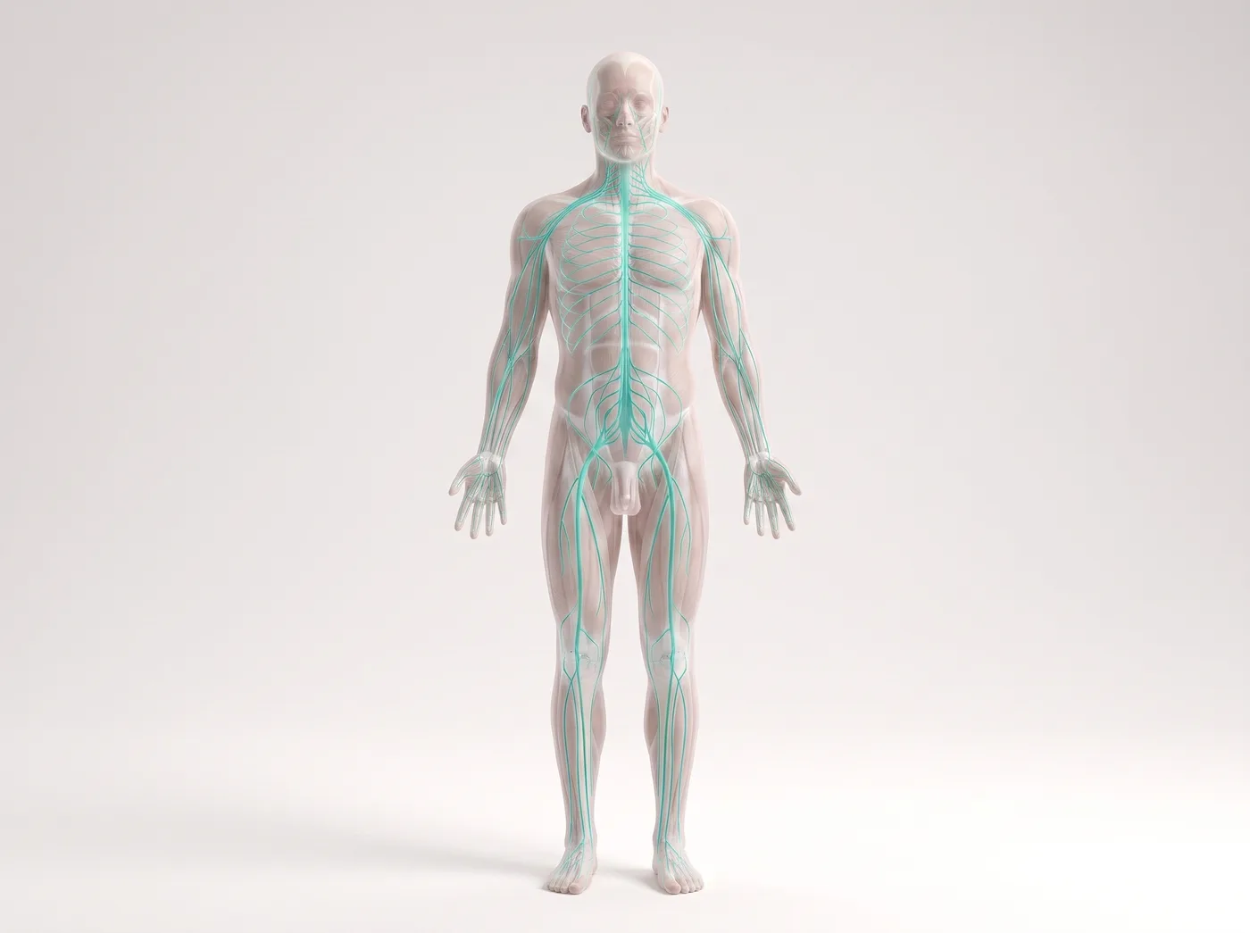
1How it works
A body-wide reset for wound-up nerve pain
2The result
You sit monitored while it runs.
What to expect: You sit monitored while it runs. In responders, relief often lasts days to weeks beyond the infusion itself — and predicts which oral medicines may help.
This page is educational material only and is not medical advice, a diagnosis, or a treatment recommendation. Illustrations are simplified artistic renderings; photos and exercise videos are AI-generated demonstrations. Evidence summaries are simplified — read the linked studies for details. Your own plan always comes from your physician. In an emergency, call 911.
 Virtual reality therapy